WB
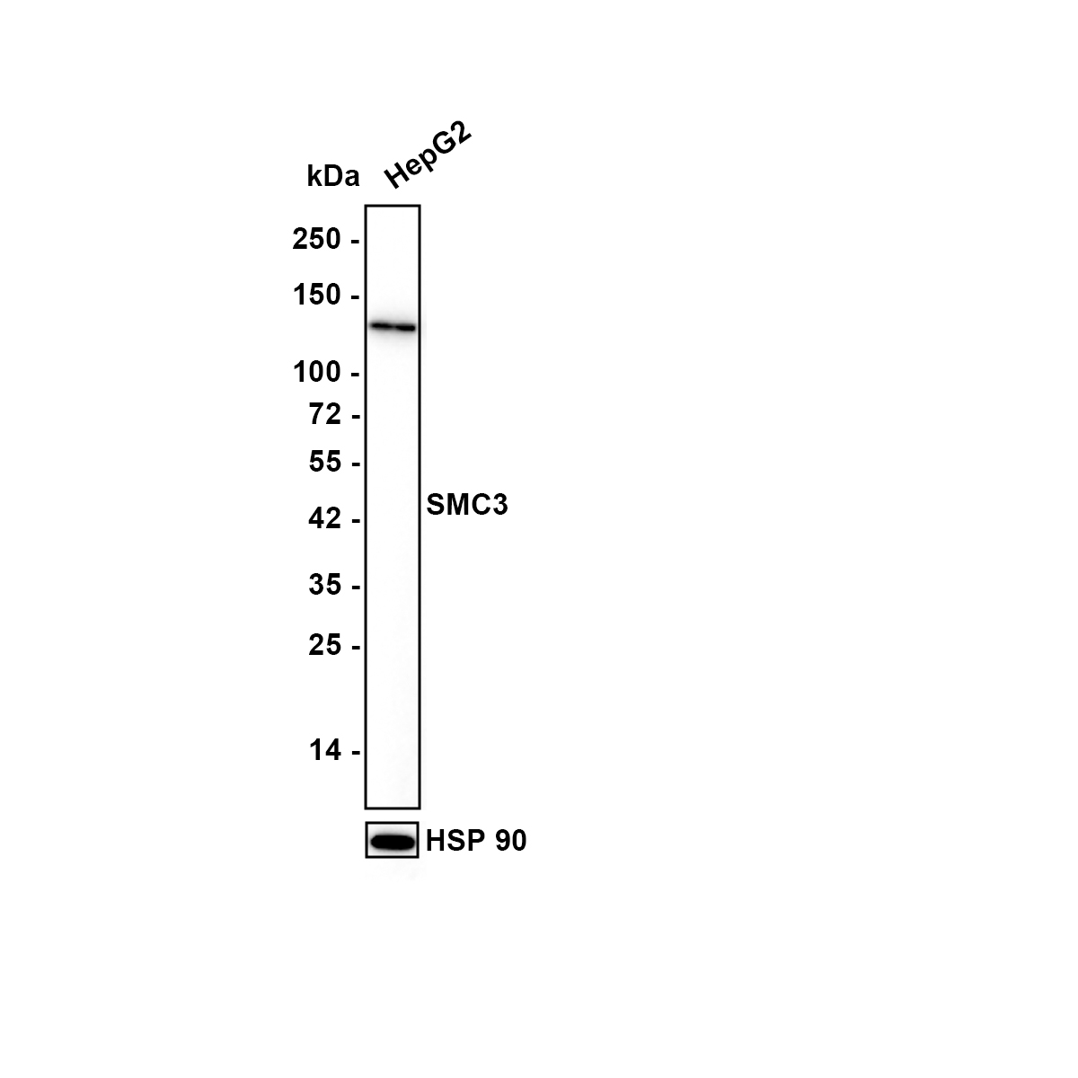
Western blot analysis of SMC3 on HepG2 lysates with Rabbit anti-SMC3 antibody at 1/5,000 dilution. Lysates/proteins at 15 µg/Lane. Exposure time: 10 seconds; Blocking: 5% NFDM/TBST, 1 hour at room temperature, Primary antibody:1/5,000 in primary antibody dilution buffer, overnight at 4 ℃. Secondary antibody: Goat anti-Rabbit IgG-HRP, 1/50,000 in 5% NFDM/TBST, 1 hour at room temperature.ICC/IF
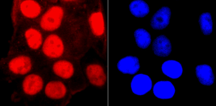
ICC staining SMC3 in Hela cells (red). The nuclear counter stain is DAPI (blue). Cells were fixed in paraformaldehyde, permeabilised with 0.25% Triton X100/PBS.ICC/IF
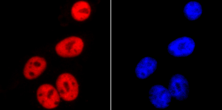
ICC staining SMC3 in HepG2 cells (red). The nuclear counter stain is DAPI (blue). Cells were fixed in paraformaldehyde, permeabilised with 0.25% Triton X100/PBS.ICC/IF
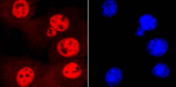
ICC staining SMC3 in NIH-3T3 cells (red). The nuclear counter stain is DAPI (blue). Cells were fixed in paraformaldehyde, permeabilised with 0.25% Triton X100/PBS.IHC
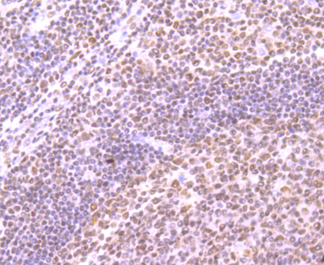
Immunohistochemical analysis of paraffin-embedded human tonsil tissue using anti-SMC3 antibody. Counter stained with hematoxylin.IHC
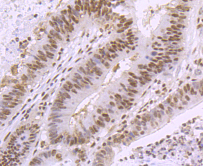
Immunohistochemical analysis of paraffin-embedded human colon cancer tissue using anti-SMC3 antibody. Counter stained with hematoxylin.IHC
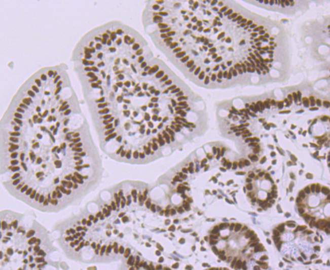
Immunohistochemical analysis of paraffin-embedded mouse colon tissue using anti-SMC3 antibody. Counter stained with hematoxylin.IHC
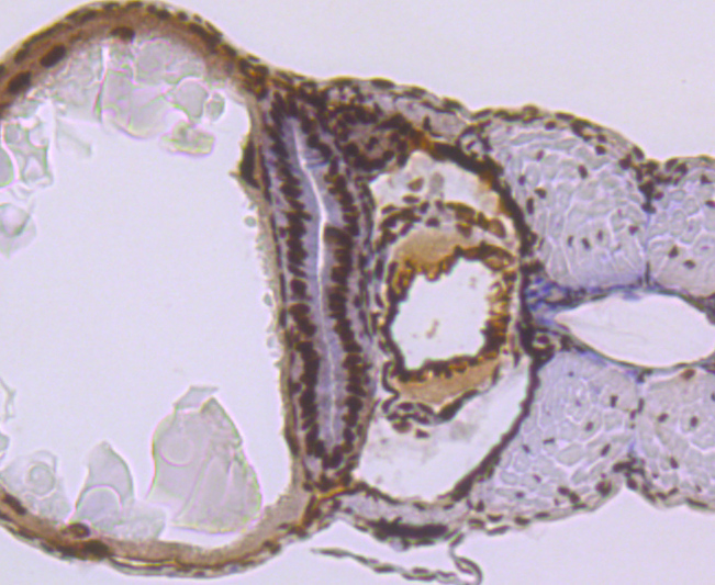
Immunohistochemical analysis of paraffin-embedded zebrafish tissue using anti-SMC3 antibody. Counter stained with hematoxylin.| Product Name | SMC3 Recombinant Rabbit Monoclonal Antibody |
|---|---|
| Antibody Type | Primary Antibodies |
| Product description | The SMC (structural maintenance of chromosomes) family of proteins form heterodimeric complexes that modulate sister chromatid cohesion and chromosome condensation for mitosis. The two distinct classes of SMC protein complexes are comprised of SMC1 (also designated SB1.8) with SMC3 (also designated HCAP for human chromosome-associated protein and Bamacan for the secreted proteoglycan), and SMC2 (also designated hCAP-E) with SMC4 (also designated hCAP-C). The SMC1/SMC3 complex is required for metaphase progression in mitotic cells and functions independently of the SMC2/SMC4 complex during the cell cycle. SMC1 is ubiqitiously expressed in various human tissues, including thymus, testis, and colon. SMC3 is expressed as a nuclear protein in the colon, but can also occur as a secreted proteoglycan expressed in testis and brain. The secreted proteoglycan contains several glycosylation sites and is thought to play a role in basement membrane physiology. |
| Immunogen | Synthetic peptide within Human SMC3 aa 1994-1217 / 1217. |
| Clonality | Monoclonal |
|---|---|
| Isotype | IgG |
| Host Species | Recombinant rabbit |
| Tested Applications | ICC/IFIHCWB |
| WB:1:1000-1:5000 IHC:1:50-1:200 ICC/IF:1:100-1:500 |
|
| Species Reactivity | HumanMouseRatZebrafish |
| Concentration | 1mg/ml |
| Purification | Protein A |
| Alternative Names | BAM antibody Bamacan antibody Basement membrane associated chondroitin proteoglycan antibody Basement membrane-associated chondroitin proteoglycan antibody BMH antibody CDLS3 antibody chondroitin sulfate proteoglycan 6 (bamacan) antibody Chondroitin sulfate proteoglycan 6 antibody Chromosome associated polypeptide antibody Chromosome-associated polypeptide antibody CSPG 6 antibody CSPG6 antibody hCAP antibody Human chromosome associated polypeptide antibody im:7142991 antibody SMC 3 antibody SMC protein 3 antibody SMC-3 antibody smc3 antibody SMC3_HUMAN antibody SMC3L1 antibody Structural maintenance of chromosome 3 antibody Structural maintenance of chromosomes 3 antibody Structural maintenance of chromosomes protein 3 antibody u:fb22e01 antibody wu:fc30d07 antibody |
|---|---|
| Molecular Weight(MW) | 142 kDa |
| Cellular Localization | Nucleus. Chromosome. |
WB
Western blot analysis of SMC3 on HepG2 lysates with Rabbit anti-SMC3 antibody at 1/5,000 dilution. Lysates/proteins at 15 µg/Lane. Exposure time: 10 seconds; Blocking: 5% NFDM/TBST, 1 hour at room temperature, Primary antibody:1/5,000 in primary antibody dilution buffer, overnight at 4 ℃. Secondary antibody: Goat anti-Rabbit IgG-HRP, 1/50,000 in 5% NFDM/TBST, 1 hour at room temperature.
ICC/IF
ICC staining SMC3 in Hela cells (red). The nuclear counter stain is DAPI (blue). Cells were fixed in paraformaldehyde, permeabilised with 0.25% Triton X100/PBS.
ICC/IF
ICC staining SMC3 in HepG2 cells (red). The nuclear counter stain is DAPI (blue). Cells were fixed in paraformaldehyde, permeabilised with 0.25% Triton X100/PBS.
ICC/IF
ICC staining SMC3 in NIH-3T3 cells (red). The nuclear counter stain is DAPI (blue). Cells were fixed in paraformaldehyde, permeabilised with 0.25% Triton X100/PBS.
IHC
Immunohistochemical analysis of paraffin-embedded human tonsil tissue using anti-SMC3 antibody. Counter stained with hematoxylin.
IHC
Immunohistochemical analysis of paraffin-embedded human colon cancer tissue using anti-SMC3 antibody. Counter stained with hematoxylin.
IHC
Immunohistochemical analysis of paraffin-embedded mouse colon tissue using anti-SMC3 antibody. Counter stained with hematoxylin.
IHC
Immunohistochemical analysis of paraffin-embedded zebrafish tissue using anti-SMC3 antibody. Counter stained with hematoxylin.| Application Notes | WB:1:1000-1:5000 IHC:1:50-1:200 ICC/IF:1:100-1:500 |
|---|
| Form | Liquid |
|---|---|
| Storage Instructions | Store at +4℃ after thawing. Aliquot store at -20℃ or -80℃. Avoid repeated freeze / thaw cycles. |
| Storage Buffer | 1*TBS (pH7.4), 1%BSA, 40%Glycerol. Preservative: 0.05% Sodium Azide. |


