Application
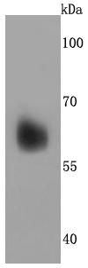
Fig1: Western blot analysis of Perilipin A on human liver cells lysates using anti-Perilipin A antibody at 1/500 dilution.Application
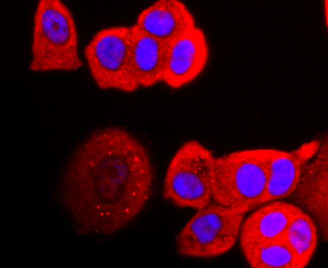
Fig2: ICC staining Perilipin A in MCF-7 cells (red). The nuclear counter stain is DAPI (blue). Cells were fixed in paraformaldehyde, permeabilised with 0.25% Triton X100/PBS.Application
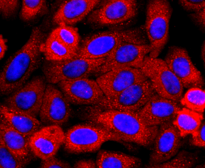
Fig3: ICC staining Perilipin A in HepG2 cells (red). The nuclear counter stain is DAPI (blue). Cells were fixed in paraformaldehyde, permeabilised with 0.25% Triton X100/PBS.Application
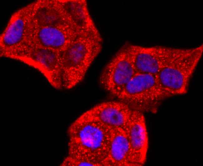
Fig4: ICC staining Perilipin A in Hela cells (red). The nuclear counter stain is DAPI (blue). Cells were fixed in paraformaldehyde, permeabilised with 0.25% Triton X100/PBS.| Product Name | Perilipin A Recombinant Rabbit Monoclonal Antibody |
|---|---|
| Antibody Type | Primary Antibodies |
| Product description | Modulator of adipocyte lipid metabolism. Coats lipid storage droplets to protect them from breakdown by hormone-sensitive lipase (HSL). Its absence may result in leanness. Plays a role in unilocular lipid droplet formation by activating CIDEC. Their interaction promotes lipid droplet enlargement and directional net neutral lipid transfer. May modulate lipolysis and triglyceride levels. |
| Immunogen | recombinant protein |
| Clonality | Monoclonal |
|---|---|
| Isotype | IgG |
| Host Species | Recombinant rabbit |
| Tested Applications | ICC/IFWB |
| WB:1:500-1:2000 ICC/IF:1:100-1:500 |
|
| Species Reactivity | HumanMouse |
| Concentration | 1mg/ml |
| Purification | Protein A |
| Alternative Names | Lipid droplet associated protein antibody PERIA antibody PERIB antibody Perilipin A antibody Perilipin antibody Perilipin B antibody PLIN antibody |
|---|---|
| Molecular Weight(MW) | 56 kDa |
| Cellular Localization | Endoplasmic reticulum, Lipid droplet. |
Application
Fig1: Western blot analysis of Perilipin A on human liver cells lysates using anti-Perilipin A antibody at 1/500 dilution.
Application
Fig2: ICC staining Perilipin A in MCF-7 cells (red). The nuclear counter stain is DAPI (blue). Cells were fixed in paraformaldehyde, permeabilised with 0.25% Triton X100/PBS.
Application
Fig3: ICC staining Perilipin A in HepG2 cells (red). The nuclear counter stain is DAPI (blue). Cells were fixed in paraformaldehyde, permeabilised with 0.25% Triton X100/PBS.
Application
Fig4: ICC staining Perilipin A in Hela cells (red). The nuclear counter stain is DAPI (blue). Cells were fixed in paraformaldehyde, permeabilised with 0.25% Triton X100/PBS.| Positive Control | MCF-7, HepG2, Hela, human liver cells lysates. |
|---|---|
| Application Notes | WB:1:500-1:2000 ICC/IF:1:100-1:500 |
| Form | Liquid |
|---|---|
| Storage Instructions | Store at +4℃ after thawing. Aliquot store at -20℃ or -80℃. Avoid repeated freeze / thaw cycles. |
| Storage Buffer | 1*TBS (pH7.4), 1%BSA, 40%Glycerol. Preservative: 0.05% Sodium Azide. |


