Application
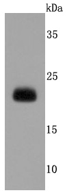
Fig1: Western blot analysis of Lipocalin-2 on A431 cells lysates using anti-Lipocalin-2 antibody at 1/500 dilution.Application
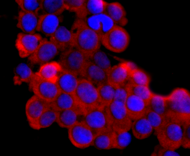
Fig2: ICC staining Lipocalin-2 in 293T cells (red). The nuclear counter stain is DAPI (blue). Cells were fixed in paraformaldehyde, permeabilised with 0.25% Triton X100/PBS.Application
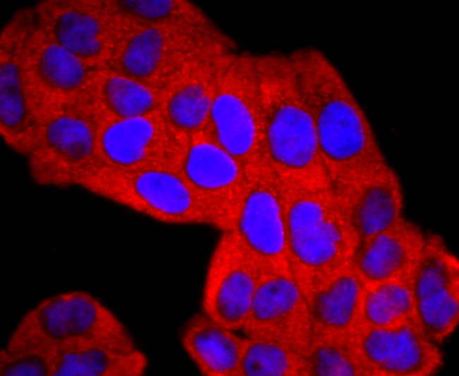
Fig3: ICC staining Lipocalin-2 in Hela cells (red). The nuclear counter stain is DAPI (blue). Cells were fixed in paraformaldehyde, permeabilised with 0.25% Triton X100/PBS.Application
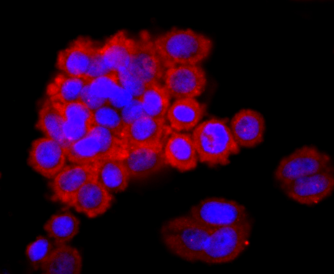
Fig4: ICC staining Lipocalin-2 in SW480 cells (red). The nuclear counter stain is DAPI (blue). Cells were fixed in paraformaldehyde, permeabilised with 0.25% Triton X100/PBS.Application
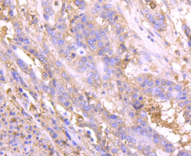
Fig5: Immunohistochemical analysis of paraffin-embedded human stomach cancer tissue using anti-Lipocalin-2 antibody. Counter stained with hematoxylin.Application
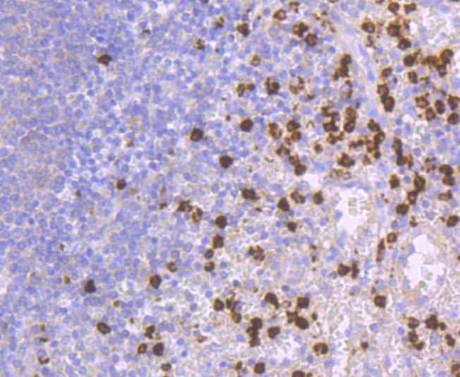
Fig6: Immunohistochemical analysis of paraffin-embedded human spleen tissue using anti-Lipocalin-2 antibody. Counter stained with hematoxylin.Application
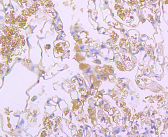
Fig7: Immunohistochemical analysis of paraffin-embedded human lung tissue using anti-Lipocalin-2 antibody. Counter stained with hematoxylin.Application
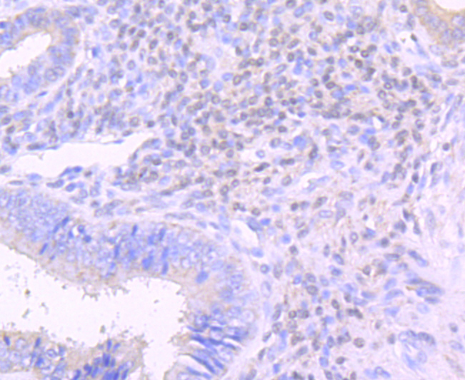
Fig8: Immunohistochemical analysis of paraffin-embedded human uterus tissue using anti-Lipocalin-2 antibody. Counter stained with hematoxylin.Application
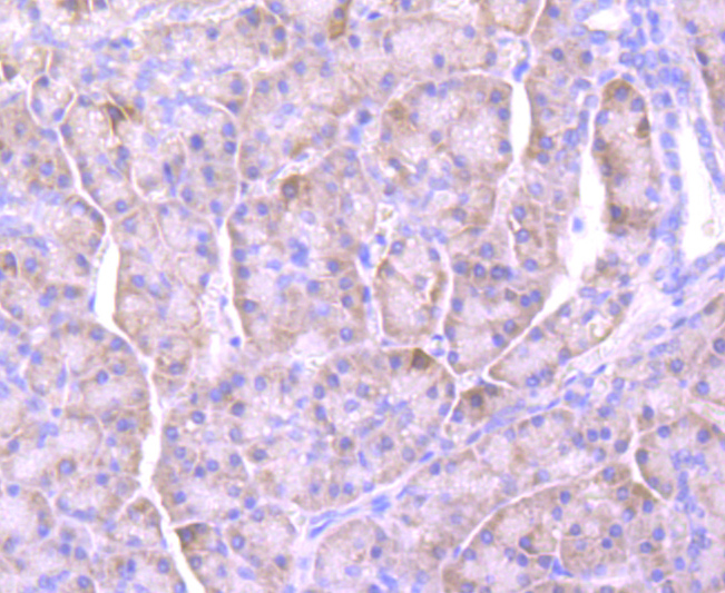
Fig9: Immunohistochemical analysis of paraffin-embedded human pancreas tissue using anti-Lipocalin-2 antibody. Counter stained with hematoxylin.| Product Name | Lipocalin-2 [JM10-67] |
|---|---|
| Antibody Type | Primary Antibodies |
| Antigen Alias | 24p3 antibody 25 kDa alpha 2 microglobulin related subunit of MMP9 antibody 25 kDa alpha-2-microglobulin-related subunit of MMP-9 antibody Alpha 2 microglobulin related protein antibody HGNC:6526 antibody HNL antibody Lcn 2 antibody Lcn2 antibody Lipocalin-2 antibody Migration stimulating factor inhibitor antibody MSFI antibody Neutrophil gelatinase associated lipocalin antibody Neutrophil gelatinase associated lipocalin precursor antibody Neutrophil gelatinase-associated lipocalin antibody NGAL antibody NGAL_HUMAN antibody Oncogene 24p3 antibody Oncogenic lipocalin 24p3 antibody p25 antibody Siderocalin antibody siderocalin LCN2 antibody SV40 induced 24P3 protein antibody Uterocalin antibody |
| Product description | In addition to the monomeric mammalian progelatinase, two additional forms of progelatinase have been identified. The shorter of these additional forms is a covalently linked, disulfide-bridged protein that heterodimerizes with a short protein; an α2-Microglobulin-related protein known as neutrophil gelatinase-associated lipocalin (NGAL), which is moderately expressed in breast and lung tissues. NGAL belongs to the lipocalin family and has a high degree of similarity with rat α2-Microglobulin-related protein and mouse protein 24p3. NGAL is able to bind a derivative of the bacterial chemotactic peptide, suggesting that it has important immuno-modulatory functions. NGAL has been described as an inflammatory protein; it is released into the circulation as a result of the inflammatory activation of leukocytes initiated by the extra-corporeal circulation. In addition, NGAL synthesis is induced in epithelial cells in inflammatory and neoplastic colorectal diseases. In conclusion, NGAL may serve as a scavenger of bacterial products to function in the anti-inflammatory process. |
| Immunogen | recombinant protein |
| Clonality | Monoclonal |
|---|---|
| Isotype | IgG |
| Host Species | Recombinant rabbit |
| Tested Applications | WBICC/IFIHC |
| WB:1:1,000-1:5,000 ICC:1:100-1:500 IHC:1:50-1:200 FC:1:50-1:100 |
|
| Species Reactivity | Human |
| Concentration | 1mg/ml |
| Alternative Names | 24p3 antibody
25 kDa alpha 2 microglobulin related subunit of MMP9 antibody 25 kDa alpha-2-microglobulin-related subunit of MMP-9 antibody Alpha 2 microglobulin related protein antibody HGNC:6526 antibody HNL antibody Lcn 2 antibody Lcn2 antibody Lipocalin-2 antibody Migration stimulating factor inhibitor antibody MSFI antibody Neutrophil gelatinase associated lipocalin antibody Neutrophil gelatinase associated lipocalin precursor antibody Neutrophil gelatinase-associated lipocalin antibody NGAL antibody NGAL_HUMAN antibody Oncogene 24p3 antibody Oncogenic lipocalin 24p3 antibody p25 antibody Siderocalin antibody siderocalin LCN2 antibody SV40 induced 24P3 protein antibody Uterocalin antibody |
|---|---|
| Molecular Weight(MW) | 23 kDa |
| Cellular Localization | Secreted. |
| SwissProt ID | P80188 |
|---|
Application
Fig1: Western blot analysis of Lipocalin-2 on A431 cells lysates using anti-Lipocalin-2 antibody at 1/500 dilution.
Application
Fig2: ICC staining Lipocalin-2 in 293T cells (red). The nuclear counter stain is DAPI (blue). Cells were fixed in paraformaldehyde, permeabilised with 0.25% Triton X100/PBS.
Application
Fig3: ICC staining Lipocalin-2 in Hela cells (red). The nuclear counter stain is DAPI (blue). Cells were fixed in paraformaldehyde, permeabilised with 0.25% Triton X100/PBS.
Application
Fig4: ICC staining Lipocalin-2 in SW480 cells (red). The nuclear counter stain is DAPI (blue). Cells were fixed in paraformaldehyde, permeabilised with 0.25% Triton X100/PBS.
Application
Fig5: Immunohistochemical analysis of paraffin-embedded human stomach cancer tissue using anti-Lipocalin-2 antibody. Counter stained with hematoxylin.
Application
Fig6: Immunohistochemical analysis of paraffin-embedded human spleen tissue using anti-Lipocalin-2 antibody. Counter stained with hematoxylin.
Application
Fig7: Immunohistochemical analysis of paraffin-embedded human lung tissue using anti-Lipocalin-2 antibody. Counter stained with hematoxylin.
Application
Fig8: Immunohistochemical analysis of paraffin-embedded human uterus tissue using anti-Lipocalin-2 antibody. Counter stained with hematoxylin.
Application
Fig9: Immunohistochemical analysis of paraffin-embedded human pancreas tissue using anti-Lipocalin-2 antibody. Counter stained with hematoxylin.| Positive Control | A431, 293T, Hela, SW480, human stomach cancer tissue, human spleen tissue, human uterus tissue, human lung tissue, human pancreas tissue. |
|---|---|
| Application Notes | WB:1:1,000-1:5,000 ICC:1:100-1:500 IHC:1:50-1:200 FC:1:50-1:100 |
| Form | Liquid |
|---|---|
| Storage Instructions | Store at +4℃ after thawing. Aliquot store at -20℃ or -80℃. Avoid repeated freeze / thaw cycles. |
| Storage Buffer | 1*TBS (pH7.4), 1%BSA, 40%Glycerol. Preservative: 0.05% Sodium Azide. |


