WB
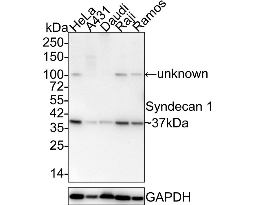
Western blot analysis of Syndecan 1 on different lysates with Rabbit anti-Syndecan 1 antibody at 1/2,000 dilution. Lane 1: HeLa cell lysate, Lane 2: A431 cell lysate, Lane 3: Daudi cell lysate, Lane 4: Raji cell lysate, Lane 5: Ramos cell lysate, Lysates/proteins at 20 µg/Lane. Exposure time: 3 minutes; 4-20% SDS-PAGE gel. Proteins were transferred to a PVDF membrane and blocked with 5% NFDM/TBST for 1 hour at room temperature. The primary antibody at 1/2,000 dilution was used in 5% NFDM/TBST at 4℃ overnight. Goat Anti-Rabbit IgG - HRP Secondary Antibody at 1:50,000 dilution was used for 1 hour at room temperature.IHC
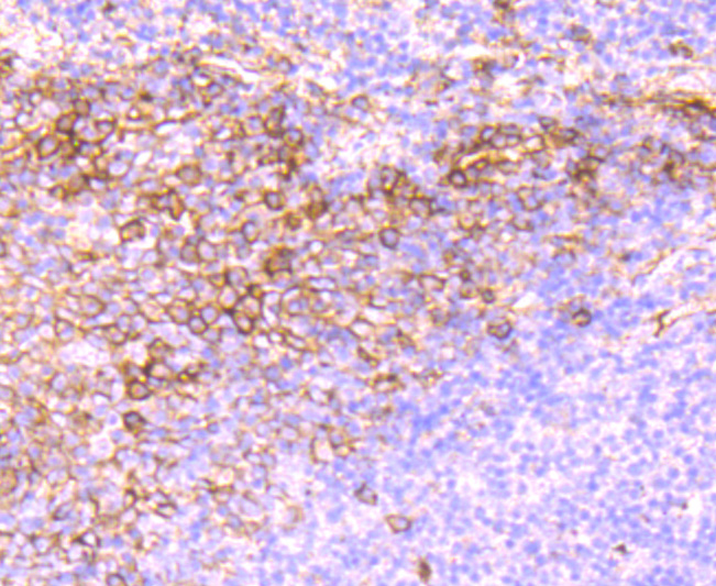
Immunohistochemical analysis of paraffin-embedded human tonsil tissue using anti-Syndecan 1 antibody. Counter stained with hematoxylin.IHC
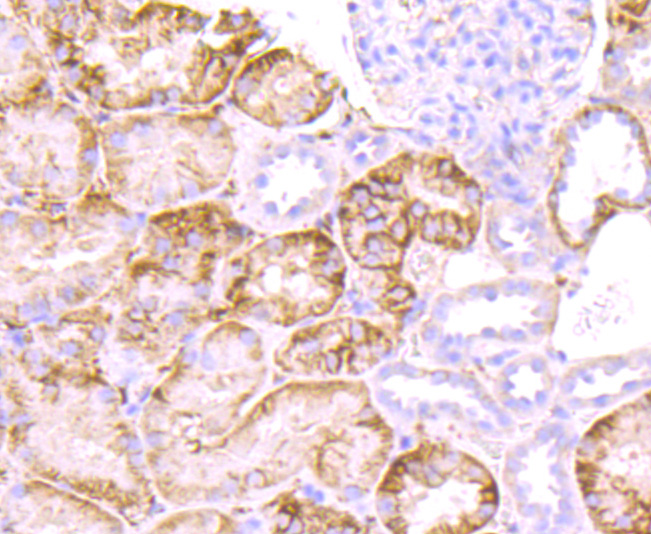
Immunohistochemical analysis of paraffin-embedded human kidney tissue using anti-Syndecan 1 antibody. Counter stained with hematoxylin.IHC
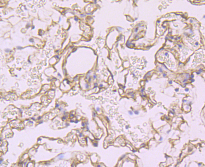
Immunohistochemical analysis of paraffin-embedded human lung tissue using anti-Syndecan 1 antibody. Counter stained with hematoxylin.IHC
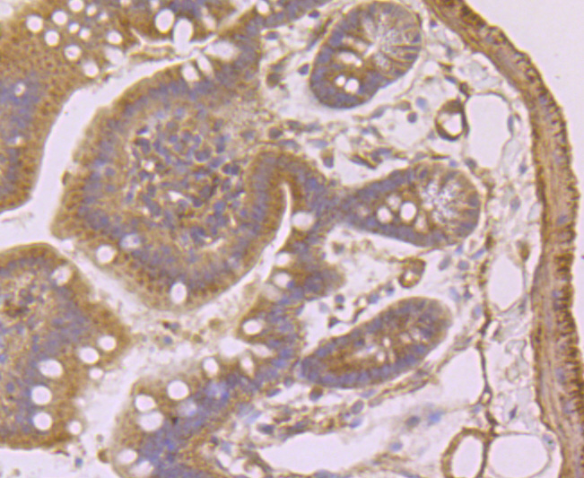
Immunohistochemical analysis of paraffin-embedded mouse colon tissue using anti-Syndecan 1 antibody. Counter stained with hematoxylin.IHC
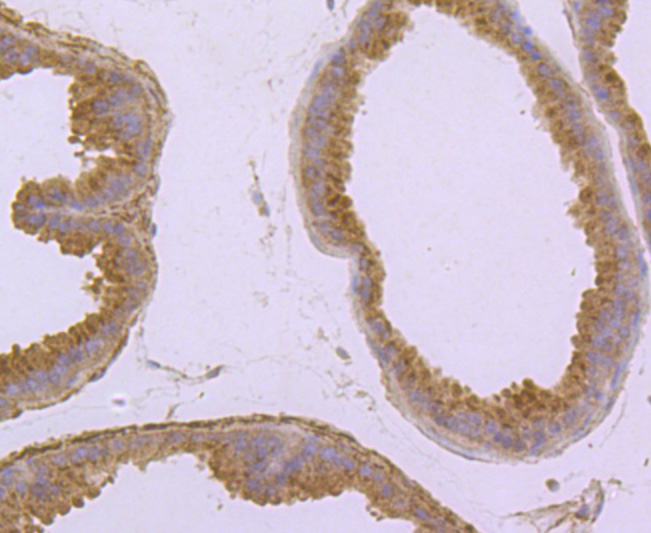
Immunohistochemical analysis of paraffin-embedded mouse prostate tissue using anti-Syndecan 1 antibody. Counter stained with hematoxylin.ICC/IF
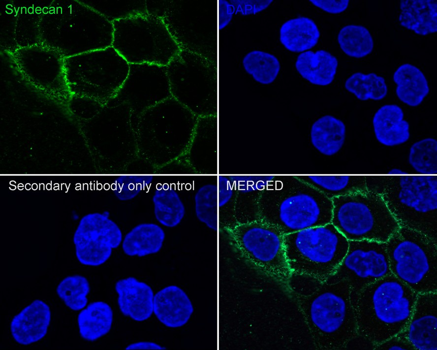
Immunocytochemistry analysis of A431 cells labeling Syndecan 1 with Rabbit anti-Syndecan 1 antibody at 1/100 dilution. Cells were fixed in 100% precooled methanol for 5 minutes at room temperature, then blocked with 1% BSA in 10% negative goat serum for 1 hour at room temperature. Cells were then incubated with Rabbit anti-Syndecan 1 antibody at 1/100 dilution in 1% BSA in PBST overnight at 4 ℃. Goat Anti-Rabbit IgG H&L (488) was used as the secondary antibody at 1/1,000 dilution. PBS instead of the primary antibody was used as the secondary antibody only control. Nuclear DNA was labelled in blue with DAPI.FC
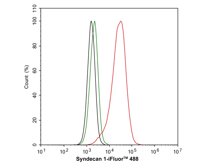
Flow cytometric analysis of HeLa cells labeling Syndecan 1. Cells were washed twice with cold PBS and resuspend. Then stained with the primary antibody (1μg/mL) (red) compared with Rabbit IgG Isotype Control (green). After incubation of the primary antibody at +4℃ for an hour, the cells were stained with a 488 conjugate-Goat anti-Rabbit IgG Secondary antibody at 1/1,000 dilution for 30 minutes at +4℃. Unlabelled sample was used as a control (cells without incubation with primary antibody; black).| Product Name | Syndecan 1 Recombinant Rabbit Monoclonal Antibody |
|---|---|
| Antibody Type | Primary Antibodies |
| Product description | Syndecan-1 (SYND1), also designated CD138, is a type I integral membrane proteoglycan that contains both chondroitin sulfate and heparan sulfate groups. It is expressed in mouse on pre-B cells, immature B cells and plasma cells. Syndecan-1 is also found on the basolateral surfaces of epithelial cells, endothelial cells of sprouting capillaries and embryonic condensing mesenchymal cells. Syndecan-1 functions as an extracellular matrix receptor which binds to collagens, Fibronectin and Thrombospondin. It has been shown to co-localize with Actin-rich filaments and may act to link the cytoskeleton to the extracellular matrix. |
| Immunogen | Recombinant protein within Human Syndecan 1 aa 13-274 / 310. |
| Clonality | Monoclonal |
|---|---|
| Isotype | IgG |
| Host Species | Rabbit |
| Tested Applications | FCICC/IFIHCWB |
| WB:1:2000 IHC:1:50-1:200 ICC/IF:1:100 FC:1:1000 |
|
| Species Reactivity | HumanMouseRat |
| Concentration | 1mg/ml |
| Purification | Protein A |
| Alternative Names | CD_antigen antibody CD138 antibody CD138 antigen antibody heparan sulfate proteoglycan fibroblast growth factor receptor antibody SDC antibody Sdc1 antibody SDC1_HUMAN antibody SYND1 antibody Syndecan 1 antibody Syndecan antibody syndecan proteoglycan 1 antibody Syndecan-1 antibody |
|---|---|
| Molecular Weight(MW) | 33 kDa(Observed band size: 37 kDa) |
| Cellular Localization | Membrane. |
WB
Western blot analysis of Syndecan 1 on different lysates with Rabbit anti-Syndecan 1 antibody at 1/2,000 dilution. Lane 1: HeLa cell lysate, Lane 2: A431 cell lysate, Lane 3: Daudi cell lysate, Lane 4: Raji cell lysate, Lane 5: Ramos cell lysate, Lysates/proteins at 20 µg/Lane. Exposure time: 3 minutes; 4-20% SDS-PAGE gel. Proteins were transferred to a PVDF membrane and blocked with 5% NFDM/TBST for 1 hour at room temperature. The primary antibody at 1/2,000 dilution was used in 5% NFDM/TBST at 4℃ overnight. Goat Anti-Rabbit IgG - HRP Secondary Antibody at 1:50,000 dilution was used for 1 hour at room temperature.
IHC
Immunohistochemical analysis of paraffin-embedded human tonsil tissue using anti-Syndecan 1 antibody. Counter stained with hematoxylin.
IHC
Immunohistochemical analysis of paraffin-embedded human kidney tissue using anti-Syndecan 1 antibody. Counter stained with hematoxylin.
IHC
Immunohistochemical analysis of paraffin-embedded human lung tissue using anti-Syndecan 1 antibody. Counter stained with hematoxylin.
IHC
Immunohistochemical analysis of paraffin-embedded mouse colon tissue using anti-Syndecan 1 antibody. Counter stained with hematoxylin.
IHC
Immunohistochemical analysis of paraffin-embedded mouse prostate tissue using anti-Syndecan 1 antibody. Counter stained with hematoxylin.
ICC/IF
Immunocytochemistry analysis of A431 cells labeling Syndecan 1 with Rabbit anti-Syndecan 1 antibody at 1/100 dilution. Cells were fixed in 100% precooled methanol for 5 minutes at room temperature, then blocked with 1% BSA in 10% negative goat serum for 1 hour at room temperature. Cells were then incubated with Rabbit anti-Syndecan 1 antibody at 1/100 dilution in 1% BSA in PBST overnight at 4 ℃. Goat Anti-Rabbit IgG H&L (488) was used as the secondary antibody at 1/1,000 dilution. PBS instead of the primary antibody was used as the secondary antibody only control. Nuclear DNA was labelled in blue with DAPI.
FC
Flow cytometric analysis of HeLa cells labeling Syndecan 1. Cells were washed twice with cold PBS and resuspend. Then stained with the primary antibody (1μg/mL) (red) compared with Rabbit IgG Isotype Control (green). After incubation of the primary antibody at +4℃ for an hour, the cells were stained with a 488 conjugate-Goat anti-Rabbit IgG Secondary antibody at 1/1,000 dilution for 30 minutes at +4℃. Unlabelled sample was used as a control (cells without incubation with primary antibody; black).| Application Notes | WB:1:2000 IHC:1:50-1:200 ICC/IF:1:100 FC:1:1000 |
|---|
| Form | Liquid |
|---|---|
| Storage Instructions | Store at +4℃ after thawing. Aliquot store at -20℃ or -80℃. Avoid repeated freeze / thaw cycles. |
| Storage Buffer | 1*TBS (pH7.4), 1%BSA, 40%Glycerol. Preservative: 0.05% Sodium Azide. |


