WB
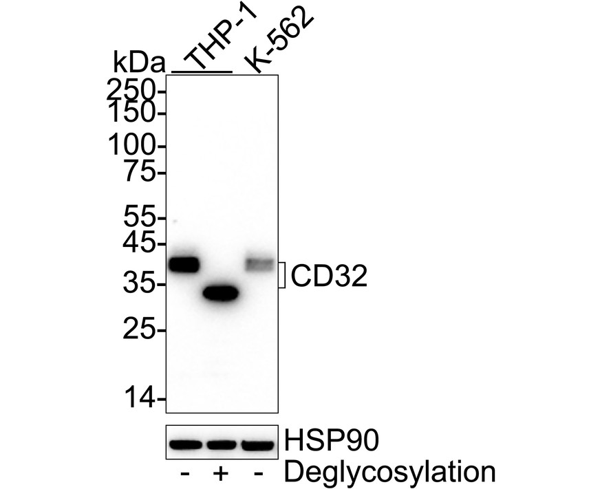
Western blot analysis of CD32 on different lysates with Rabbit anti-CD32 antibody at 1/2,000 dilution. Lane 1: THP-1 cell lysate, Lane 2: THP-1 cell lysate treated with deglycosylation, Lane 3: K-562 cell lysate, Lysates/proteins at 20 µg/Lane. Exposure time: 3 minutes; 4-20% SDS-PAGE gel. Proteins were transferred to a PVDF membrane and blocked with 5% NFDM/TBST for 1 hour at room temperature. The primary antibody at 1/2,000 dilution was used in 5% NFDM/TBST at 4℃ overnight. Goat Anti-Rabbit IgG - HRP Secondary Antibody at 1/50,000 dilution was used for 1 hour at room temperature.IHC
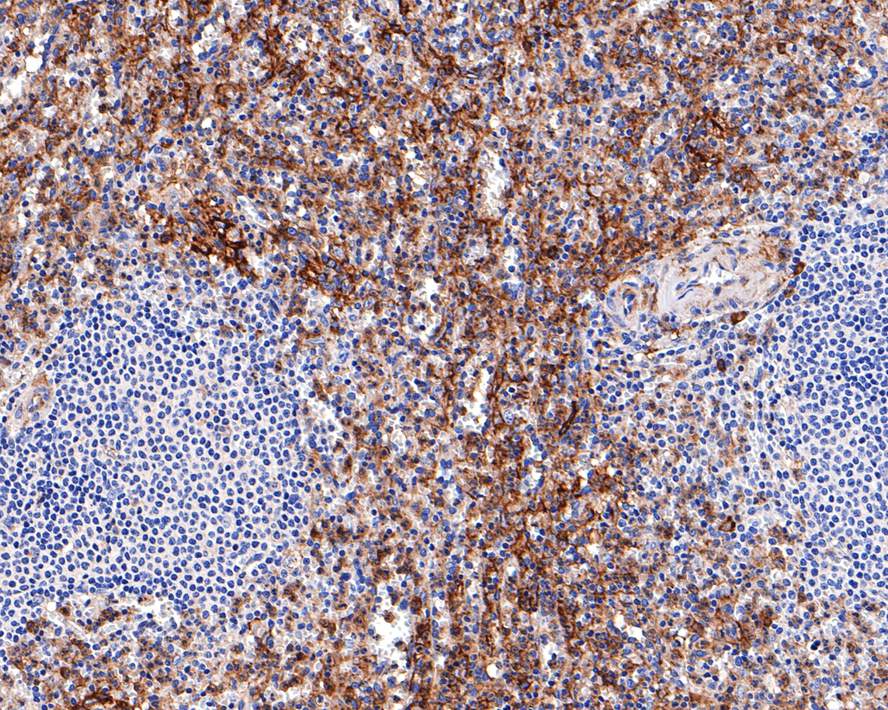
Immunohistochemical analysis of paraffin-embedded human spleen tissue with Rabbit anti-CD32 antibody at 1/400 dilution. The section was pre-treated using heat mediated antigen retrieval with Tris-EDTA buffer (pH 9.0) for 20 minutes. The tissues were blocked in 1% BSA for 20 minutes at room temperature, washed with ddH2O and PBS, and then probed with the primary antibody at 1/400 dilution for 1 hour at room temperature. The detection was performed using an HRP conjugated compact polymer system. DAB was used as the chromogen. Tissues were counterstained with hematoxylin and mounted with DPX.ICC/IF
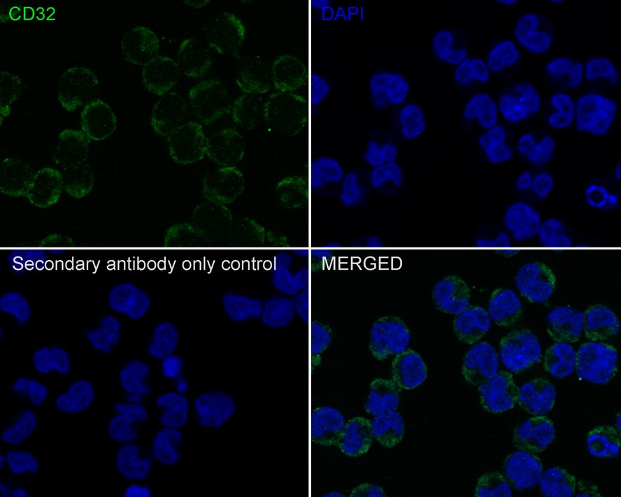
Immunocytochemistry analysis of K-562 cells labeling CD32 with Rabbit anti-CD32 antibody at 1/100 dilution. Cells were fixed in 80% precooled methanol for 5 minutes at room temperature, then blocked with 1% BSA in 10% negative goat serum for 1 hour at room temperature. Cells were then incubated with Rabbit anti-CD32 antibody at 1/100 dilution in 1% BSA in PBST overnight at 4 ℃. Goat Anti-Rabbit IgG H&L (488) was used as the secondary antibody at 1/1,000 dilution. PBS instead of the primary antibody was used as the secondary antibody only control. Nuclear DNA was labelled in blue with DAPI.FC
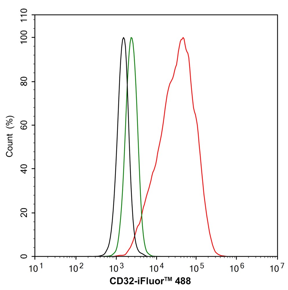
Flow cytometric analysis of K-562 cells labeling CD32. Cells were fixed and permeabilized. Then stained with the primary antibody (1/1,000) (red) compared with Rabbit IgG Isotype Control (green). After incubation of the primary antibody at +4℃ for an hour, the cells were stained with a 488 conjugate-Goat anti-Rabbit IgG Secondary antibody at 1/1,000 dilution for 30 minutes at +4℃. Unlabelled sample was used as a control (cells without incubation with primary antibody; black).IP
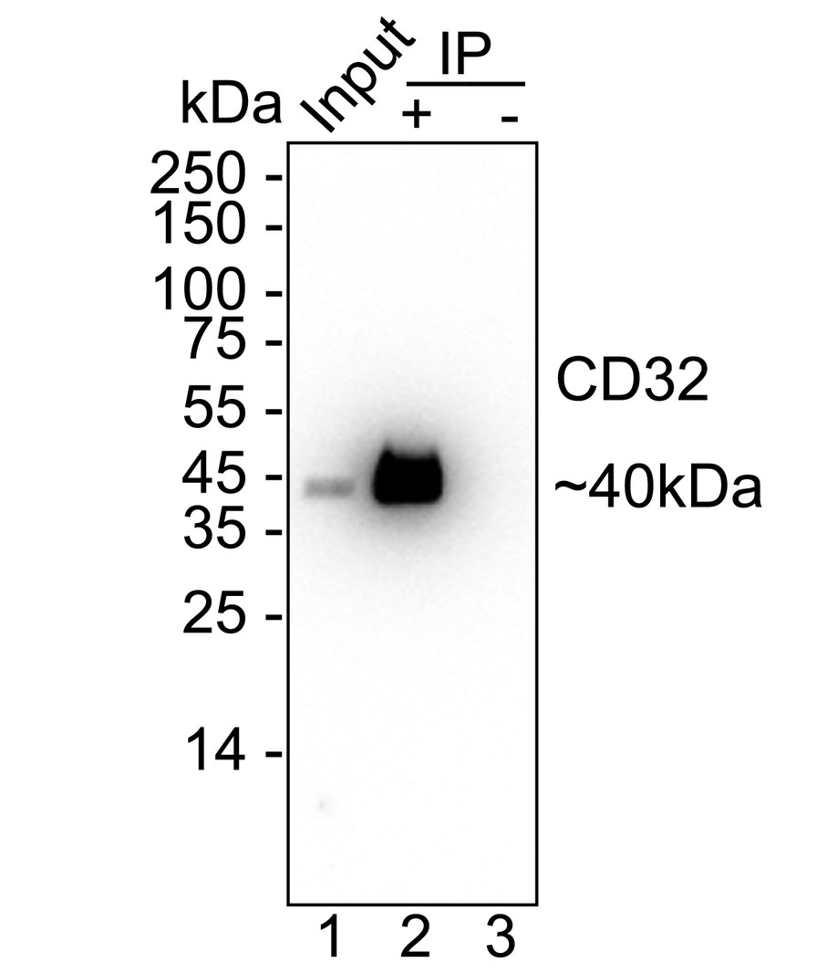
CD32 was immunoprecipitated from 0.2 mg THP-1 cell lysate with Rabbit anti-CD32 antibody at 2 µg/10 µl beads. Western blot was performed from the immunoprecipitate using Rabbit anti-CD32 antibody at 1/2,000 dilution. HRP Conjugated Anti-Rabbit IgG for IP Nano-secondary antibody at 1/5,000 dilution was used for 1 hour at room temperature. Lane 1: THP-1 cell lysate (input), Lane 2: Rabbit anti-CD32 antibody IP in THP-1 cell lysate, Lane 3: Rabbit IgG instead of Rabbit anti-CD32 antibody in THP-1 cell lysate, Blocking/Dilution buffer: 5% NFDM/TBST, Exposure time: 1 minute 7 seconds.| Product Name | CD32 Recombinant Rabbit Monoclonal Antibody |
|---|---|
| Antibody Type | Primary Antibodies |
| Product description | CD32 (also designated Fc gamma RII) is a low affinity receptor for the Fc fragment of aggregated IgG (1,2). CD32 is responsible for the clearance of immunocomplexes by macrophages and also plays an important role in the regulation of antibody production by B cells (1-4). IgG can noncooperatively bind either one or two highly glycosylated CD32 molecules, and this binding delivers a negative signal for B cells (1,2,5). CD32 exists as several isoforms that are produced by alternative splicing of three distinct genes, A, B, and C (2,6). These isoforms are designated FcgRIIA, FcgRIIB1, FcgRIIB3, and FcgRIIC (1,2,6). All isoforms are present on monocytes, placental trophoblasts and endothelial cells (1,6). In addition, the FcgRIIB forms are present on B lymphocytes, and the FcgRIIA and FcgRIIC forms are found on neutrophils (1,6). |
| Immunogen | Recombinant protein within Human CD32 aa 23-274 / 310. |
| Clonality | Monoclonal |
|---|---|
| Isotype | IgG |
| Host Species | Recombinant rabbit |
| Tested Applications | FCICC/IFIHCIPWB |
| WB:1:1000-1:5000 IHC1:400ICC/IF:1:100 FC:1:1000 IP:1-2μg/sample |
|
| Species Reactivity | Human |
| Concentration | 1mg/ml |
| Purification | Protein A |
| Alternative Names | CD32 antibody
CD32A antibody
CD32B antibody
CD32C antibody
CDw32 antibody
Fc fragment of IgG low affinity IIa receptor antibody
Fc fragment of IgG low affinity IIa receptor for (CD32) antibody
Fc fragment of IgG low affinity IIb receptor antibody
Fc fragment of IgG low affinity IIb receptor for (CD32) antibody
Fc fragment of IgG low affinity IIc receptor antibody
Fc fragment of IgG low affinity IIc receptor for (CD32) antibody
Fc fragment of IgG low affinity II receptor for (CD32) antibody Fc fragment of IgG low affinity IIa receptor (CD32) antibody Fc fragment of IgG low affinity IIb receptor (CD32) antibody Fc gamma receptor IIB antibody Fc gamma receptor IIC antibody Fc gamma RII a antibody Fc gamma RII b antibody Fc gamma RII c antibody Fc gamma RIIb antibody Fc gamma RIIc antibody Fc receptor IgG low affinity IIb antibody Fc-gamma RII-a antibody Fc-gamma RII-b antibody Fc-gamma RII-c antibody Fc-gamma-RIIa antibody FC-gamma-RIIB antibody Fc-GAMMA-RIIC antibody Fc[g]RII antibody FCG2 antibody FCG2A_HUMAN antibody FcGR antibody FCGR2 antibody FCGR2A antibody FCGR2A1 antibody FCGR2B antibody FCGR2C antibody Fcgr3 antibody Fcgr3a antibody FcgRII antibody Fcr-2 antibody Fcr-3 antibody FcRII a antibody FCRII antibody FcRII b antibody FcRII c antibody FcRII-a antibody FcRIIC antibody IGFR2 antibody IgG Fc receptor II a antibody IgG Fc receptor II b antibody IgG Fc receptor II beta antibody IgG Fc receptor II c antibody IgG Fc receptor II-a antibody IgG Fc receptor II-b antibody IgG Fc receptor II-c antibody Immunoglobulin G Fc receptor II antibody Immunoglobulin G Fc receptor II c antibody Low affinity immunoglobulin gamma Fc region receptor II antibody Low affinity immunoglobulin gamma Fc region receptor II-a antibody Low affinity immunoglobulin gamma Fc region receptor II-b antibody Low affinity immunoglobulin gamma Fc region receptor II-c antibody Ly-17 antibody Ly-m20 antibody LyM-1 antibody Lymphocyte antigen 17 antibody |
|---|---|
| Molecular Weight(MW) | 35 kDa(Observed band size: 40/31 kDa) |
| Cellular Localization | Cell membrane. |
| SwissProt ID | P12318 |
|---|
WB
Western blot analysis of CD32 on different lysates with Rabbit anti-CD32 antibody at 1/2,000 dilution. Lane 1: THP-1 cell lysate, Lane 2: THP-1 cell lysate treated with deglycosylation, Lane 3: K-562 cell lysate, Lysates/proteins at 20 µg/Lane. Exposure time: 3 minutes; 4-20% SDS-PAGE gel. Proteins were transferred to a PVDF membrane and blocked with 5% NFDM/TBST for 1 hour at room temperature. The primary antibody at 1/2,000 dilution was used in 5% NFDM/TBST at 4℃ overnight. Goat Anti-Rabbit IgG - HRP Secondary Antibody at 1/50,000 dilution was used for 1 hour at room temperature.
IHC
Immunohistochemical analysis of paraffin-embedded human spleen tissue with Rabbit anti-CD32 antibody at 1/400 dilution. The section was pre-treated using heat mediated antigen retrieval with Tris-EDTA buffer (pH 9.0) for 20 minutes. The tissues were blocked in 1% BSA for 20 minutes at room temperature, washed with ddH2O and PBS, and then probed with the primary antibody at 1/400 dilution for 1 hour at room temperature. The detection was performed using an HRP conjugated compact polymer system. DAB was used as the chromogen. Tissues were counterstained with hematoxylin and mounted with DPX.
ICC/IF
Immunocytochemistry analysis of K-562 cells labeling CD32 with Rabbit anti-CD32 antibody at 1/100 dilution. Cells were fixed in 80% precooled methanol for 5 minutes at room temperature, then blocked with 1% BSA in 10% negative goat serum for 1 hour at room temperature. Cells were then incubated with Rabbit anti-CD32 antibody at 1/100 dilution in 1% BSA in PBST overnight at 4 ℃. Goat Anti-Rabbit IgG H&L (488) was used as the secondary antibody at 1/1,000 dilution. PBS instead of the primary antibody was used as the secondary antibody only control. Nuclear DNA was labelled in blue with DAPI.
FC
Flow cytometric analysis of K-562 cells labeling CD32. Cells were fixed and permeabilized. Then stained with the primary antibody (1/1,000) (red) compared with Rabbit IgG Isotype Control (green). After incubation of the primary antibody at +4℃ for an hour, the cells were stained with a 488 conjugate-Goat anti-Rabbit IgG Secondary antibody at 1/1,000 dilution for 30 minutes at +4℃. Unlabelled sample was used as a control (cells without incubation with primary antibody; black).
IP
CD32 was immunoprecipitated from 0.2 mg THP-1 cell lysate with Rabbit anti-CD32 antibody at 2 µg/10 µl beads. Western blot was performed from the immunoprecipitate using Rabbit anti-CD32 antibody at 1/2,000 dilution. HRP Conjugated Anti-Rabbit IgG for IP Nano-secondary antibody at 1/5,000 dilution was used for 1 hour at room temperature. Lane 1: THP-1 cell lysate (input), Lane 2: Rabbit anti-CD32 antibody IP in THP-1 cell lysate, Lane 3: Rabbit IgG instead of Rabbit anti-CD32 antibody in THP-1 cell lysate, Blocking/Dilution buffer: 5% NFDM/TBST, Exposure time: 1 minute 7 seconds.| Positive Control | Human lung, human tonsil tissue, human spleen tissue, human pancreas tissue. |
|---|---|
| Application Notes | WB:1:1000-1:5000 IHC1:400ICC/IF:1:100 FC:1:1000 IP:1-2μg/sample |
| Form | Liquid |
|---|---|
| Storage Instructions | Store at +4℃ after thawing. Aliquot store at -20℃ or -80℃. Avoid repeated freeze / thaw cycles. |
| Storage Buffer | 1*TBS (pH7.4), 1%BSA, 40%Glycerol. Preservative: 0.05% Sodium Azide. |


