Application
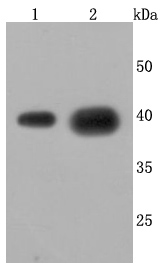
Fig1: Western blot analysis of Cathepsin L/V/K/H on different cells lysates using anti-Cathepsin L/V/K/H antibody at 1/1,000 dilution. Positive control: Lane 1: HepG2 Lane 2: A549Application
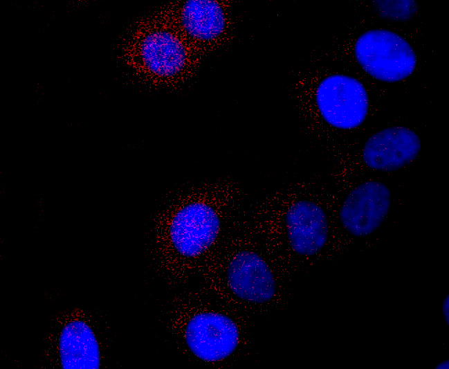
Fig2: ICC staining Cathepsin L/V/K/H in HepG2 cells (red). The nuclear counter stain is DAPI (blue). Cells were fixed in paraformaldehyde, permeabilised with 0.25% Triton X100/PBS.Application
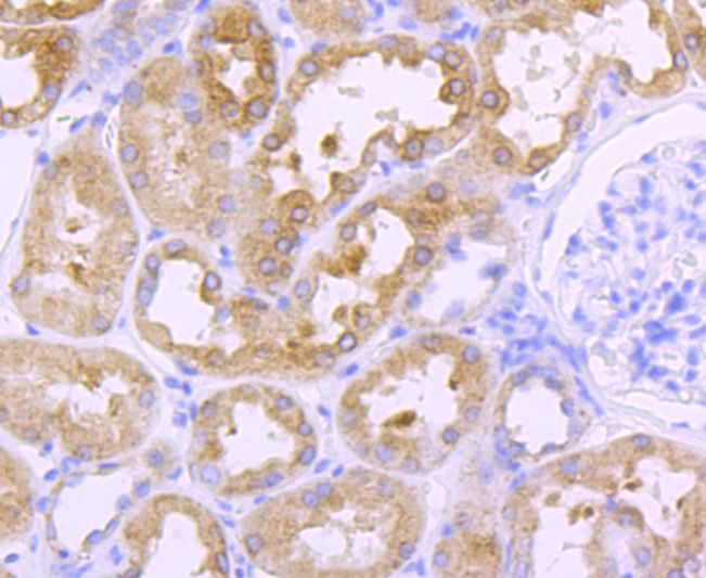
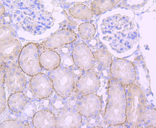
Fig3: Immunohistochemical analysis of paraffin-embedded human kidney tissue using anti-Cathepsin L/V/K/H antibody. Counter stained with hematoxylin.Application
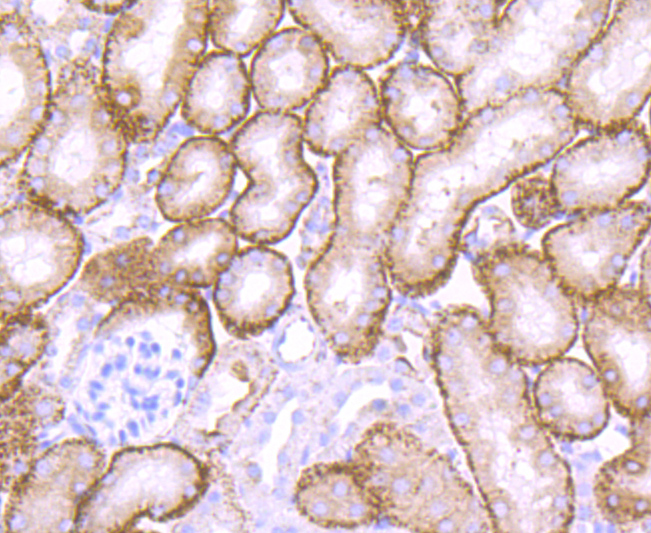
Fig4: Immunohistochemical analysis of paraffin-embedded mouse kidney tissue using anti-Cathepsin L/V/K/H antibody. Counter stained with hematoxylin.Application
Fig5: Immunohistochemical analysis of paraffin-embedded rat kidney tissue using anti-Cathepsin L/V/K/H antibody. Counter stained with hematoxylin.Application
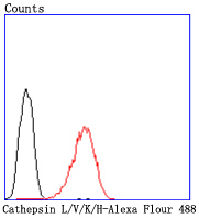
Fig6: Flow cytometric analysis of HepG2 cells with Cathepsin L/V/K/H antibody at 1/50 dilution (red) compared with an unlabelled control (cells without incubation with primary antibody; black). Alexa Fluor 488-conjugated goat anti rabbit IgG was used as the secondary antibody.| Product Name | Cathepsin LVKH [JM10-78] |
|---|---|
| Antibody Type | Primary Antibodies |
| Product description | The cathepsin family of proteolytic enzymes contains several diverse classes of proteases. The cysteine protease class comprises cathepsins B, L, H, K, S, and O. The aspartyl protease class is composed of cathepsins D and E. Cathepsin G is in the serine protease class. Most cathepsins are lysosomal and each is involved in cellular metabolism, participating in various events such as peptide biosynthesis and protein degradation. Cathepsin L (also designated major excreted protein, MEP or CATL) is a member of the peptidase C1 family and has been identified as a protein that is most closely related to cathepsin H. It is a lysosomal cysteine proteinase that mediates intracellular protein catabolism for collagen, elastin and ?-1 protease inhibitor. Cathepsin L is a dimer composed of disulfide-linked heavy and light chains, both produced from a single protein precursor. At least two transcript variants encoding the same protein have been found for this gene. Transformed mouse fibroblasts stimulated by growth factors or tumor promoters secrete a form of cathepsin L. |
| Immunogen | recombinant protein |
| Clonality | Monoclonal |
|---|---|
| Isotype | IgG |
| Host Species | Recombinant rabbit |
| Tested Applications | WBICC/IFIHCFC |
| WB:1:1,000-1:5,000 ICC:1:50-1:100 IHC:1:50-1:200 FC:1:50-1:100 |
|
| Species Reactivity | HumanMouseRat |
| Concentration | 1mg/ml |
| Molecular Weight(MW) | 38 kDa |
|---|---|
| Cellular Localization | Lysosome. |
Application
Fig1: Western blot analysis of Cathepsin L/V/K/H on different cells lysates using anti-Cathepsin L/V/K/H antibody at 1/1,000 dilution. Positive control: Lane 1: HepG2 Lane 2: A549
Application
Fig2: ICC staining Cathepsin L/V/K/H in HepG2 cells (red). The nuclear counter stain is DAPI (blue). Cells were fixed in paraformaldehyde, permeabilised with 0.25% Triton X100/PBS.
Application
Fig3: Immunohistochemical analysis of paraffin-embedded human kidney tissue using anti-Cathepsin L/V/K/H antibody. Counter stained with hematoxylin.
Application
Fig4: Immunohistochemical analysis of paraffin-embedded mouse kidney tissue using anti-Cathepsin L/V/K/H antibody. Counter stained with hematoxylin.
Application
Fig5: Immunohistochemical analysis of paraffin-embedded rat kidney tissue using anti-Cathepsin L/V/K/H antibody. Counter stained with hematoxylin.
Application
Fig6: Flow cytometric analysis of HepG2 cells with Cathepsin L/V/K/H antibody at 1/50 dilution (red) compared with an unlabelled control (cells without incubation with primary antibody; black). Alexa Fluor 488-conjugated goat anti rabbit IgG was used as the secondary antibody.| Positive Control | HepG2, A549, human kidney tissue, mouse kidney tissue, rat kidney tissue. |
|---|---|
| Application Notes | WB:1:1,000-1:5,000 ICC:1:50-1:100 IHC:1:50-1:200 FC:1:50-1:100 |
| Form | Liquid |
|---|---|
| Storage Instructions | Store at +4℃ after thawing. Aliquot store at -20℃ or -80℃. Avoid repeated freeze / thaw cycles. |
| Storage Buffer | 1*TBS (pH7.4), 1%BSA, 40%Glycerol. Preservative: 0.05% Sodium Azide. |


