Application
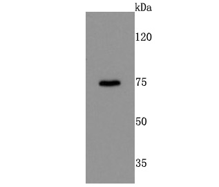
Fig1: Western blot analysis of DRD1 on mouse kidney cells lysates using anti-DRD1 antibody at 1/500 dilution.Application
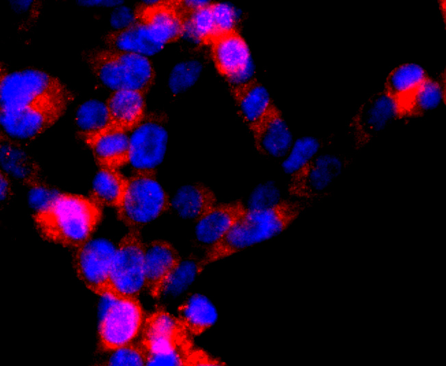
Fig2: ICC staining DRD1 in 293T cells (red). The nuclear counter stain is DAPI (blue). Cells were fixed in paraformaldehyde, permeabilised with 0.25% Triton X100/PBS.Application
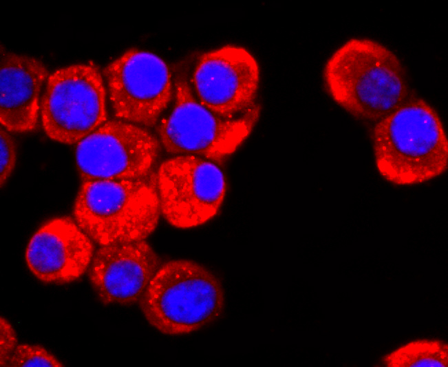
Fig3: ICC staining DRD1 in N2A cells (red). The nuclear counter stain is DAPI (blue). Cells were fixed in paraformaldehyde, permeabilised with 0.25% Triton X100/PBS.Application
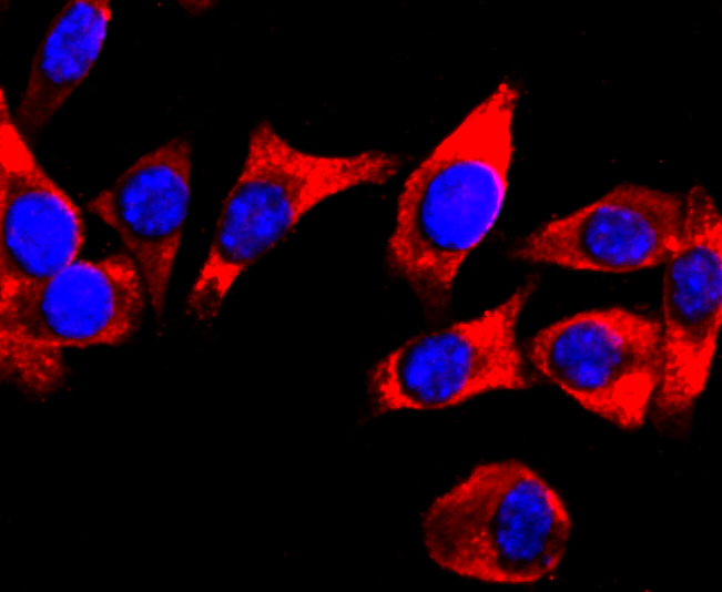
Fig4: ICC staining DRD1 in SH-SY5Y cells (red). The nuclear counter stain is DAPI (blue). Cells were fixed in paraformaldehyde, permeabilised with 0.25% Triton X100/PBS.Application
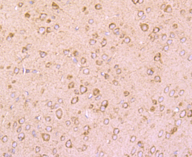
Fig5: Immunohistochemical analysis of paraffin-embedded mouse brain tissue using anti-DRD1 antibody. Counter stained with hematoxylin.Application
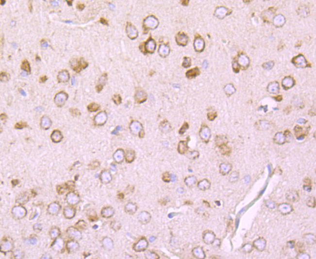
Fig6: Immunohistochemical analysis of paraffin-embedded rat brain tissue using anti-DRD1 antibody. Counter stained with hematoxylin.| Product Name | DRD1 [JM10-93] |
|---|---|
| Antibody Type | Primary Antibodies |
| Antigen Alias | D(1A) dopamine receptor antibody D1A dopamine receptor antibody DADR antibody Dopamine D1 receptor antibody dopamine receptor D1 antibody DR D1 antibody DR D1A antibody DRD 1 antibody DRD 1A antibody DRD1 antibody DRD1_HUMAN antibody DRD1A antibody |
| Product description | The members of the G protein coupled receptor family are distinguished by their slow transmitting response to ligand binding. These transmembrane proteins include the adrenergic, serotonin and dopamine receptors. The effect of the signaling molecule can be excitatory or inhibitory depending on the type of receptor to which it binds. β-adrenergic receptor binds to adrenaline activates adenylyl cyclase, while α2-adrenergic receptor binds to adrenaline inhibits adenylyl cyclase. The dopamine receptors are divided into two classes, D1 and D2, which differ in their functional characteristics in that D1 receptors stimulate adenylyl cyclase while D2 receptors inhibit adenylyl cyclase activity. Five different subtypes of dopamine receptor have been described to date. D1DR and D5DR belong to the D1 subclass, while D2DR, D3DR and D4DR belong to the D2 subclass. |
| Immunogen | recombinant protein |
| Clonality | Monoclonal |
|---|---|
| Isotype | IgG |
| Host Species | Recombinant rabbit |
| Tested Applications | WBICC/IFIHC |
| WB:1:500-1:1,000 ICC:1:100-1:500 IHC:1:50-1:200 |
|
| Species Reactivity | HumanMouseRat |
| Concentration | 1mg/ml |
| Alternative Names | D(1A) dopamine receptor antibody D1A dopamine receptor antibody DADR antibody Dopamine D1 receptor antibody dopamine receptor D1 antibody DR D1 antibody DR D1A antibody DRD 1 antibody DRD 1A antibody DRD1 antibody DRD1_HUMAN antibody DRD1A antibody |
|---|---|
| Molecular Weight(MW) | 75 kDa |
| Cellular Localization | Cell membrane. Endoplasmic reticulum membrane. |
Application
Fig1: Western blot analysis of DRD1 on mouse kidney cells lysates using anti-DRD1 antibody at 1/500 dilution.
Application
Fig2: ICC staining DRD1 in 293T cells (red). The nuclear counter stain is DAPI (blue). Cells were fixed in paraformaldehyde, permeabilised with 0.25% Triton X100/PBS.
Application
Fig3: ICC staining DRD1 in N2A cells (red). The nuclear counter stain is DAPI (blue). Cells were fixed in paraformaldehyde, permeabilised with 0.25% Triton X100/PBS.
Application
Fig4: ICC staining DRD1 in SH-SY5Y cells (red). The nuclear counter stain is DAPI (blue). Cells were fixed in paraformaldehyde, permeabilised with 0.25% Triton X100/PBS.
Application
Fig5: Immunohistochemical analysis of paraffin-embedded mouse brain tissue using anti-DRD1 antibody. Counter stained with hematoxylin.
Application
Fig6: Immunohistochemical analysis of paraffin-embedded rat brain tissue using anti-DRD1 antibody. Counter stained with hematoxylin.| Positive Control | 293T, N2A, SH-SY5Y, mouse kidney, mouse brain tissue, rat brain tissue. |
|---|---|
| Application Notes | WB:1:500-1:1,000 ICC:1:100-1:500 IHC:1:50-1:200 |
| Form | Liquid |
|---|---|
| Storage Instructions | Store at +4℃ after thawing. Aliquot store at -20℃ or -80℃. Avoid repeated freeze / thaw cycles. |
| Storage Buffer | 1*TBS (pH7.4), 1%BSA, 40%Glycerol. Preservative: 0.05% Sodium Azide. |


