Application
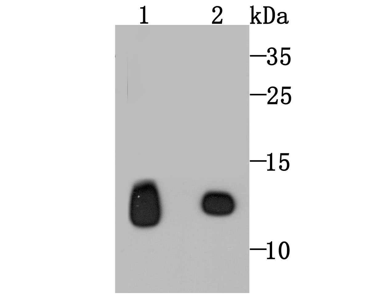
Fig1: Western blot analysis of HBG1/2 on human placenta (1) and human brain (2) tissue lysates using anti-HBG1/2 antibody at 1/500 dilution.Application
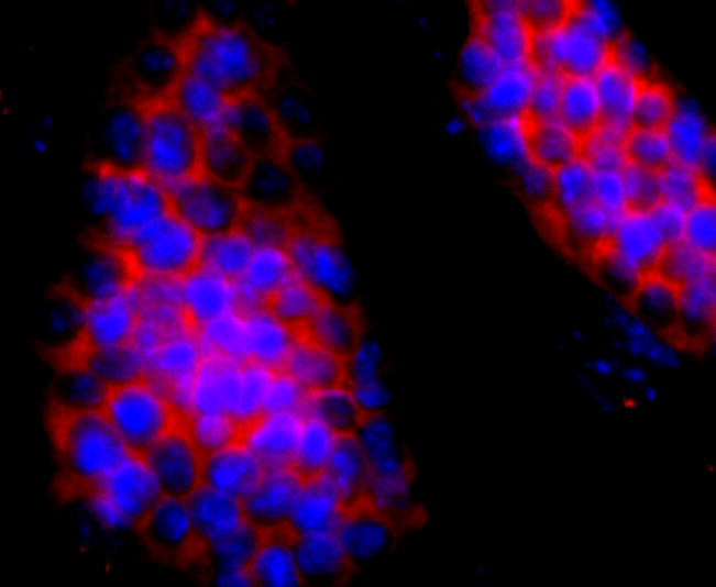
Fig2: ICC staining HBG1/2 in D3 cells (red). The nuclear counter stain is DAPI (blue). Cells were fixed in paraformaldehyde, permeabilised with 0.25% Triton X100/PBS.Application
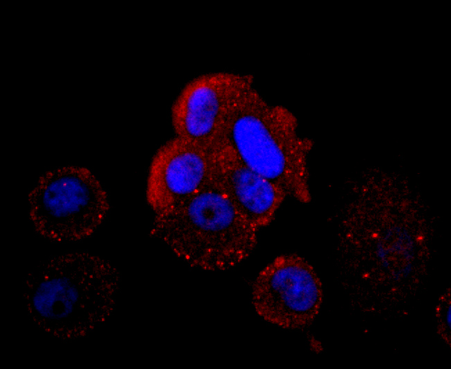
Fig3: ICC staining HBG1/2 in MCF-7 cells (red). The nuclear counter stain is DAPI (blue). Cells were fixed in paraformaldehyde, permeabilised with 0.25% Triton X100/PBS.Application
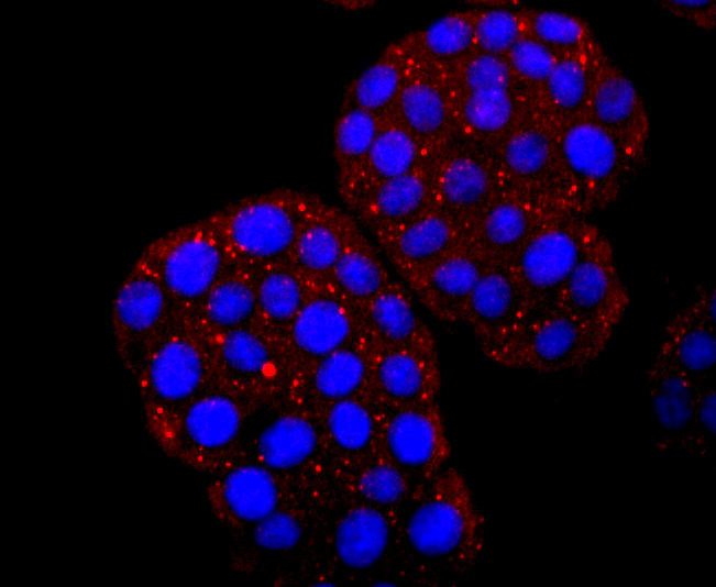
Fig4: ICC staining HBG1/2 in PC-12 cells (red). The nuclear counter stain is DAPI (blue). Cells were fixed in paraformaldehyde, permeabilised with 0.25% Triton X100/PBS.Application
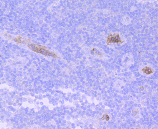
Fig5: Immunohistochemical analysis of paraffin-embedded human tonsil tissue using anti-HBG1/2 antibody. Counter stained with hematoxylin.Application
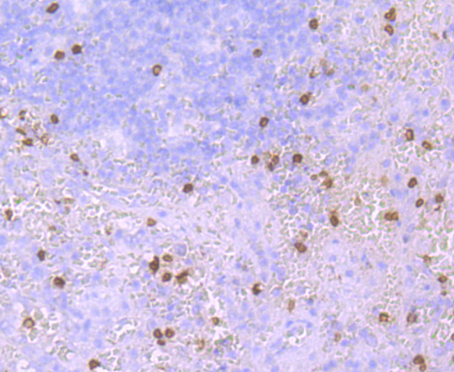
Fig6: Immunohistochemical analysis of paraffin-embedded human spleen tissue using anti-HBG1/2 antibody. Counter stained with hematoxylin.| Product Name | Hemoglobin subunit gamma 1and2 [JM84-10] |
|---|---|
| Antibody Type | Primary Antibodies |
| Product description | Hemoglobin (Hgb) is coupled to four iron-binding, methene-linked tetrapyrrole rings (heme). The α (16p13.3; 5′-ζ-pseudoz-pseudo α2-pseudo α1-α2-α1-?1-3′) and β (11p15.5) globin loci determine the basic hemoglobin structure. The globin portion of hemoglobin consists of two α chains and two β chains arranged in pairs forming a tetramer. Each of the four globin chains covalently associates with a heme group. The bonds between α and β chains are weaker than between similar globin chains, thereby forming a cleavage plane that is important for oxygen binding and release. High affinity for oxygen occurs upon relaxation of the α1-β2 cleavage plane. When the two α1-β2 interfaces are closely bound, hemoglobin has a low affinity for oxygen. Hb A, which contains two α chains plus two β chains, comprises 97% of total circulating hemoglobin. The remaining 3% of total circulating hemoglobin is comprised of Hb A-2, which consists of two α chains plus two δ chains, and fetal hemoglobin (Hb F), which consists of two α chains together with two γ chains. |
| Immunogen | recombinant protein |
| Clonality | Monoclonal |
|---|---|
| Isotype | IgG |
| Host Species | Recombinant rabbit |
| Tested Applications | WBICC/IFIHC |
| WB:1:1,000-1:5,000 ICC:1:50-1:200 IHC:1:50-1:200 FC:1:50-1:100: |
|
| Species Reactivity | Human |
| Concentration | 1mg/ml |
| Molecular Weight(MW) | 16 kDa |
|---|---|
| Cellular Localization | Cytosol, hemoglobin complex. |
Application
Fig1: Western blot analysis of HBG1/2 on human placenta (1) and human brain (2) tissue lysates using anti-HBG1/2 antibody at 1/500 dilution.
Application
Fig2: ICC staining HBG1/2 in D3 cells (red). The nuclear counter stain is DAPI (blue). Cells were fixed in paraformaldehyde, permeabilised with 0.25% Triton X100/PBS.
Application
Fig3: ICC staining HBG1/2 in MCF-7 cells (red). The nuclear counter stain is DAPI (blue). Cells were fixed in paraformaldehyde, permeabilised with 0.25% Triton X100/PBS.
Application
Fig4: ICC staining HBG1/2 in PC-12 cells (red). The nuclear counter stain is DAPI (blue). Cells were fixed in paraformaldehyde, permeabilised with 0.25% Triton X100/PBS.
Application
Fig5: Immunohistochemical analysis of paraffin-embedded human tonsil tissue using anti-HBG1/2 antibody. Counter stained with hematoxylin.
Application
Fig6: Immunohistochemical analysis of paraffin-embedded human spleen tissue using anti-HBG1/2 antibody. Counter stained with hematoxylin.| Positive Control | D3, MCF-7, PC-12, human placenta cells lysates. |
|---|---|
| Application Notes | WB:1:1,000-1:5,000 ICC:1:50-1:200 IHC:1:50-1:200 FC:1:50-1:100: |
| Form | Liquid |
|---|---|
| Storage Instructions | Store at +4℃ after thawing. Aliquot store at -20℃ or -80℃. Avoid repeated freeze / thaw cycles. |
| Storage Buffer | 1*TBS (pH7.4), 1%BSA, 40%Glycerol. Preservative: 0.05% Sodium Azide. |


