WB
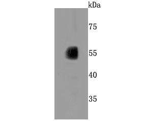
Western blot analysis of CYP2E1 on human liver cells lysates using anti-CYP2E1 antibody at 1/500 dilution.WB
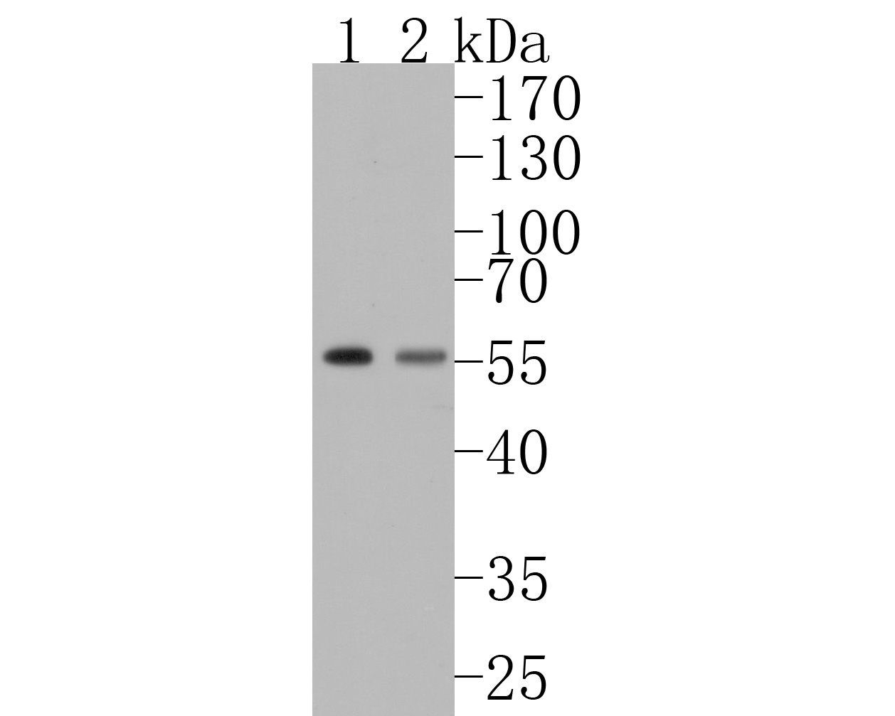
Western blot analysis of CYP2E1 on different lysates. Proteins were transferred to a PVDF membrane and blocked with 5% BSA in PBS for 1 hour at room temperature. The primary antibody (1/500) was used in 5% BSA at room temperature for 2 hours. Goat Anti-Rabbit IgG - HRP Secondary Antibody at 1:200,000 dilution was used for 1 hour at room temperature. Positive control: Lane 1: Mouse liver tissue lysate, Lane 2: Rat liver tissue lysate.IHC
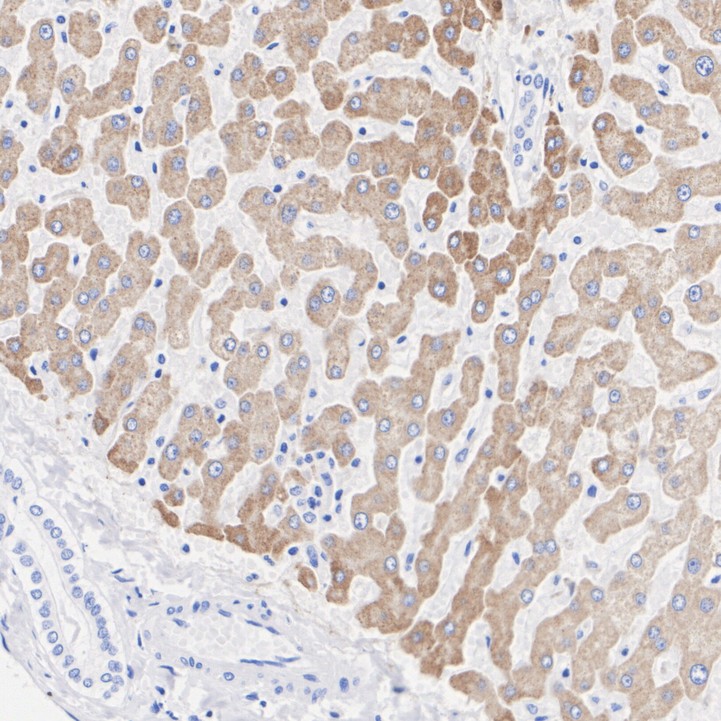
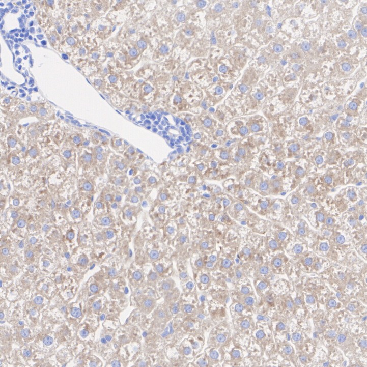
Immunohistochemical analysis of paraffin-embedded human liver tissue with Rabbit anti-CYP2E1 antibody at 1/200 dilution. The section was pre-treated using heat mediated antigen retrieval with Tris-EDTA buffer (pH 9.0) for 20 minutes. The tissues were blocked in 1% BSA for 20 minutes at room temperature, washed with ddH2O and PBS, and then probed with the primary antibody at 1/200 dilution for 1 hour at room temperature. The detection was performed using an HRP conjugated compact polymer system. DAB was used as the chromogen. Tissues were counterstained with hematoxylin and mounted with DPX.IHC
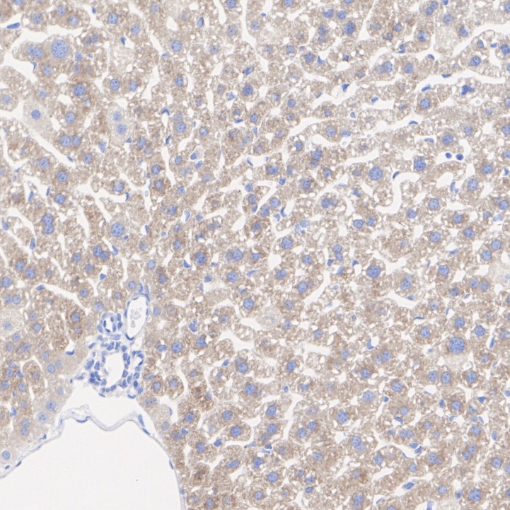
Immunohistochemical analysis of paraffin-embedded mouse liver tissue with Rabbit anti-CYP2E1 antibody at 1/200 dilution. The section was pre-treated using heat mediated antigen retrieval with Tris-EDTA buffer (pH 9.0) for 20 minutes. The tissues were blocked in 1% BSA for 20 minutes at room temperature, washed with ddH2O and PBS, and then probed with the primary antibody at 1/200 dilution for 1 hour at room temperature. The detection was performed using an HRP conjugated compact polymer system. DAB was used as the chromogen. Tissues were counterstained with hematoxylin and mounted with DPX.IHC
Immunohistochemical analysis of paraffin-embedded rat liver tissue with Rabbit anti-CYP2E1 antibody at 1/200 dilution. The section was pre-treated using heat mediated antigen retrieval with Tris-EDTA buffer (pH 9.0) for 20 minutes. The tissues were blocked in 1% BSA for 20 minutes at room temperature, washed with ddH2O and PBS, and then probed with the primary antibody at 1/200 dilution for 1 hour at room temperature. The detection was performed using an HRP conjugated compact polymer system. DAB was used as the chromogen. Tissues were counterstained with hematoxylin and mounted with DPX.| Product Name | CYP2E1 Recombinant Rabbit Monoclonal Antibody |
|---|---|
| Antibody Type | Primary Antibodies |
| Product description | Metabolizes several precarcinogens, drugs, and solvents to reactive metabolites. Inactivates a number of drugs and xenobiotics and also bioactivates many xenobiotic substrates to their hepatotoxic or carcinogenic forms. |
| Immunogen | Synthetic peptide within Human CYP2E1 aa 71-114 / 439. |
| Clonality | monoclonal |
|---|---|
| Isotype | IgG |
| Host Species | Recombinant rabbit |
| Tested Applications | IHCWB |
| WB:1:1000-1:5000 IHC:1:50-1:200 |
|
| Species Reactivity | HumanMouseRat |
| Concentration | 1mg/ |
| Purification | Protein A |
| Alternative Names | 4-nitrophenol 2-hydroxylase antibody
CP2E1_HUMAN antibody
CPE1 antibody
CYP2E antibody
Cyp2e1 antibody
CYPIIE1 antibody
Cytochrome P450 2E1 antibody
Cytochrome P450 isozyme 3A antibody
Cytochrome P450 LM3A antibody
Cytochrome P450 family 2 subfamily e polypeptide 1 antibody cytochrome P450 subfamily IIE (ethanol-inducible) polypeptide 1 antibody Cytochrome P450 subfamily IIE antibody Cytochrome P450-ALC antibody Cytochrome P450-J antibody Cytochrome P450RLM6 antibody EC 1.14.13.- antibody EC 1.14.13.n7 antibody Ethanol-inducible P450 antibody Flavoprotein-linked monooxygenase antibody MGC133529 antibody Microsomal monooxygenase antibody OTTHUMP00000020813 antibody OTTHUMP00000046571 antibody P450 ALC antibody P450-J antibody P450C2E antibody P450J antibody P450RLM6 antibody Xenobiotic monooxygenase antibody |
|---|---|
| Molecular Weight(MW) | 57kDa |
| Cellular Localization | Endoplasmic reticulum, Membrane, Microsome, Mitochondrion, Mitochondrion inner membrane. |
WB
Western blot analysis of CYP2E1 on human liver cells lysates using anti-CYP2E1 antibody at 1/500 dilution.
WB
Western blot analysis of CYP2E1 on different lysates. Proteins were transferred to a PVDF membrane and blocked with 5% BSA in PBS for 1 hour at room temperature. The primary antibody (1/500) was used in 5% BSA at room temperature for 2 hours. Goat Anti-Rabbit IgG - HRP Secondary Antibody at 1:200,000 dilution was used for 1 hour at room temperature. Positive control: Lane 1: Mouse liver tissue lysate, Lane 2: Rat liver tissue lysate.
IHC
Immunohistochemical analysis of paraffin-embedded human liver tissue with Rabbit anti-CYP2E1 antibody at 1/200 dilution. The section was pre-treated using heat mediated antigen retrieval with Tris-EDTA buffer (pH 9.0) for 20 minutes. The tissues were blocked in 1% BSA for 20 minutes at room temperature, washed with ddH2O and PBS, and then probed with the primary antibody at 1/200 dilution for 1 hour at room temperature. The detection was performed using an HRP conjugated compact polymer system. DAB was used as the chromogen. Tissues were counterstained with hematoxylin and mounted with DPX.
IHC
Immunohistochemical analysis of paraffin-embedded mouse liver tissue with Rabbit anti-CYP2E1 antibody at 1/200 dilution. The section was pre-treated using heat mediated antigen retrieval with Tris-EDTA buffer (pH 9.0) for 20 minutes. The tissues were blocked in 1% BSA for 20 minutes at room temperature, washed with ddH2O and PBS, and then probed with the primary antibody at 1/200 dilution for 1 hour at room temperature. The detection was performed using an HRP conjugated compact polymer system. DAB was used as the chromogen. Tissues were counterstained with hematoxylin and mounted with DPX.
IHC
Immunohistochemical analysis of paraffin-embedded rat liver tissue with Rabbit anti-CYP2E1 antibody at 1/200 dilution. The section was pre-treated using heat mediated antigen retrieval with Tris-EDTA buffer (pH 9.0) for 20 minutes. The tissues were blocked in 1% BSA for 20 minutes at room temperature, washed with ddH2O and PBS, and then probed with the primary antibody at 1/200 dilution for 1 hour at room temperature. The detection was performed using an HRP conjugated compact polymer system. DAB was used as the chromogen. Tissues were counterstained with hematoxylin and mounted with DPX.| Application Notes | WB:1:1000-1:5000 IHC:1:50-1:200 |
|---|
| Form | Liquid |
|---|---|
| Storage Instructions | Store at +4℃ after thawing. Aliquot store at -20℃ or -80℃. Avoid repeated freeze / thaw cycles. |
| Storage Buffer | 1*TBS (pH7.4), 1%BSA, 40%Glycerol. Preservative: 0.05% Sodium Azide. |


