Application
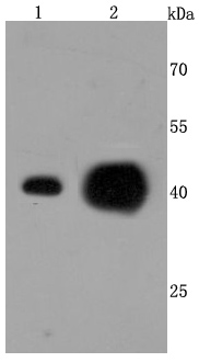
Fig1: Western blot analysis of ATP5A1 on different cells lysates using anti-ATP5A1 antibody at 1/500 dilution. Positive control: Line 1: Hela Line 2: MCF-7Application
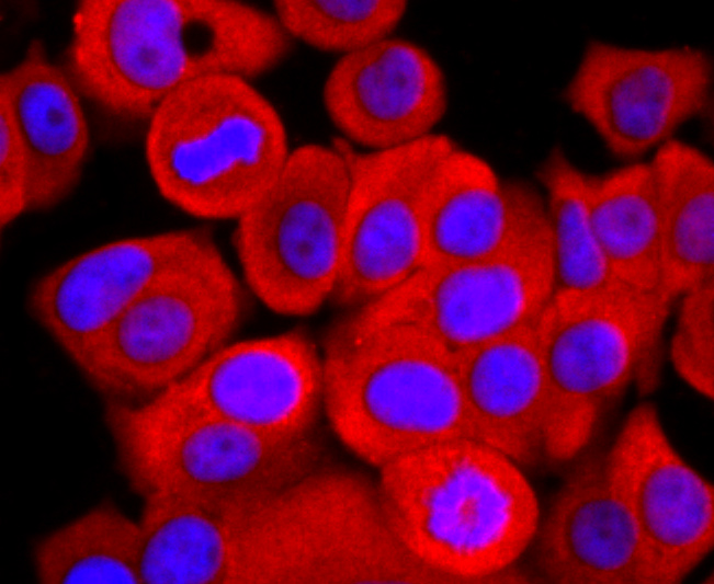
Fig2: ICC staining ATP5A1 in MCF-7 cells (red). The nuclear counter stain is DAPI (blue). Cells were fixed in paraformaldehyde, permeabilised with 0.25% Triton X100/PBS.Application
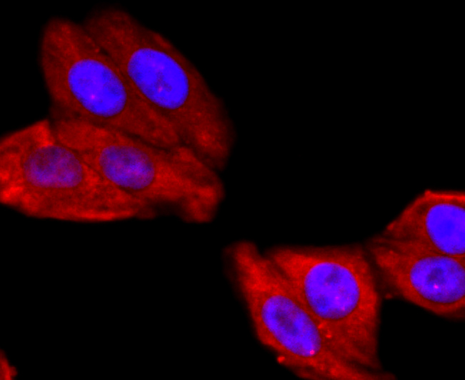
Fig3: ICC staining ATP5A1 in HepG2 cells (red). The nuclear counter stain is DAPI (blue). Cells were fixed in paraformaldehyde, permeabilised with 0.25% Triton X100/PBS.Application
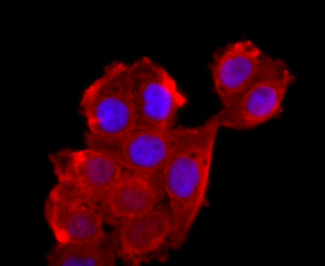
Fig4: ICC staining ATP5A1 in Hela cells (red). The nuclear counter stain is DAPI (blue). Cells were fixed in paraformaldehyde, permeabilised with 0.25% Triton X100/PBS.Application
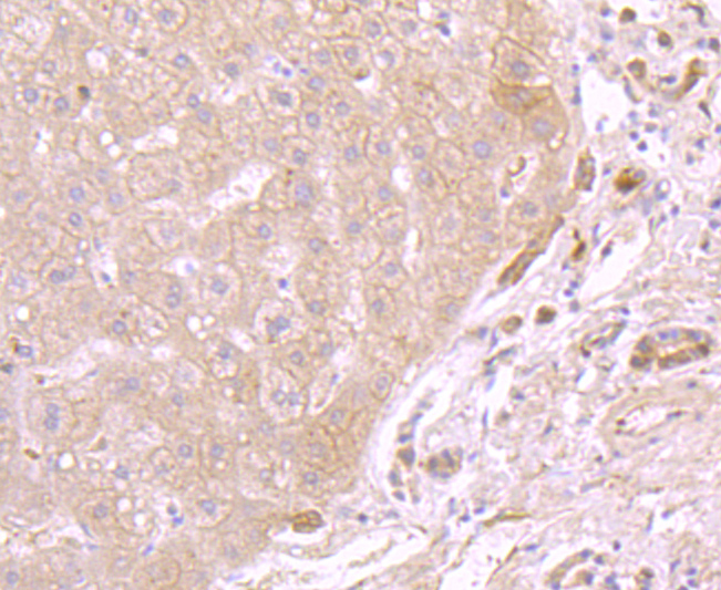
Fig5: Immunohistochemical analysis of paraffin-embedded human liver tissue using anti-ATP5A1 antibody. Counter stained with hematoxylin.Application
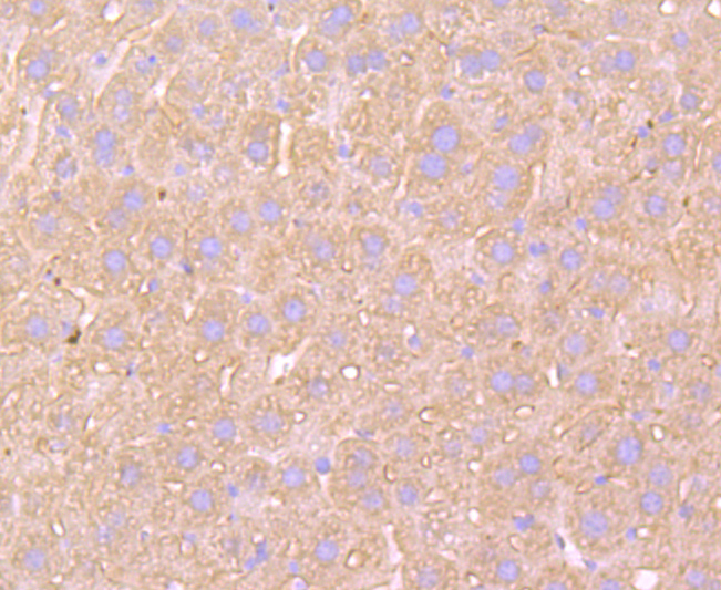
Fig6: Immunohistochemical analysis of paraffin-embedded mouse liver tissue using anti-ATP5A1 antibody. Counter stained with hematoxylin.Application
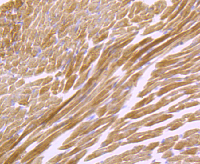
Fig7: Immunohistochemical analysis of paraffin-embedded mouse heart muscle tissue using anti-ATP5A1 antibody. Counter stained with hematoxylin.Application
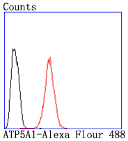
Fig8: Flow cytometric analysis of Hela cells with ATP5A1 antibody at 1/50 dilution (red) compared with an unlabelled control (cells without incubation with primary antibody; black). Alexa Fluor 488-conjugated goat anti rabbit IgG was used as the secondary antibody.| Product Name | ATP5A1 [JM110-04] |
|---|---|
| Antibody Type | Primary Antibodies |
| Antigen Alias | ATP synthase alpha chain, mitochondrial antibody ATP synthase subunit alpha antibody ATP synthase subunit alpha mitochondrial antibody ATP synthase, H+ transporting, mitochondrial F1 complex, alpha subunit 1, cardiac muscle antibody ATP synthase, H+ transporting, mitochondrial F1 complex, alpha subunit, 1 antibody ATP synthase, H+ transporting, mitochondrial F1 complex, alpha subunit, isoform 1, cardiac muscle antibody ATP synthase, H+ transporting, mitochondrial F1 complex, alpha subunit, isoform 2, non-cardiac muscle-like 2 antibody ATP sythase (F1 ATPase) alpha subunit antibody ATP5A antibody Atp5a1 antibody ATP5AL2 antibody ATPA_HUMAN antibody ATPM antibody Epididymis secretory sperm binding protein Li 123m antibody hATP1 antibody HEL-S-123m antibody MC5DN4 antibody mitochondrial antibody Mitochondrial ATP synthetase antibody Mitochondrial ATP synthetase oligomycin resistant antibody Modifier of Min 2 mouse homolog antibody Modifier of Min 2, mouse, homolog of antibody MOM2 antibody OMR antibody ORM antibody OTTHUMP00000163475 antibody |
| Product description | Mitochondrial ATP synthases (ATPases) transduce the energy contained in membrane electrochemical proton gradients into the energy required for synthesis of high-energy phosphate bonds. ATPases contain two linked complexes: F1, the hydrophilic catalytic core; and F0, the membrane-embedded protein channel. F1 consists of three α chains and three β chains, which are weakly homologous, as well as one γ chain, one δ chain and one e chain. F0 consists of three subunits: a, b and c. The α chain of F1 is a regulatory subunit that contains 509 amino acids. Mitochondrial ATPase α chain (ATP5A) localizes to the mitochondria and catalyzes ATP synthesis. |
| Immunogen | recombinant protein |
| Clonality | Monoclonal |
|---|---|
| Isotype | IgG |
| Host Species | Recombinant rabbit |
| Tested Applications | WBICC/IFIHCFC |
| WB:1:1,000-1:5,000 ICC:1:100-1:500 IHC:1:50-1:200 FC:1:50-1:100 |
|
| Species Reactivity | HumanMouseRat |
| Concentration | 1mg/ml |
| Alternative Names | ATP synthase alpha chain mitochondrial antibody ATP synthase subunit alpha antibody ATP synthase subunit alpha mitochondrial antibody ATP synthase H+ transporting mitochondrial F1 complex alpha subunit 1 cardiac muscle antibody ATP synthase H+ transporting mitochondrial F1 complex alpha subunit 1 antibody ATP synthase H+ transporting mitochondrial F1 complex alpha subunit isoform 1 cardiac muscle antibody ATP synthase H+ transporting mitochondrial F1 complex alpha subunit isoform 2 non-cardiac muscle-like 2 antibody ATP sythase (F1 ATPase) alpha subunit antibody ATP5A antibody Atp5a1 antibody ATP5AL2 antibody ATPA_HUMAN antibody ATPM antibody Epididymis secretory sperm binding protein Li 123m antibody hATP1 antibody HEL-S-123m antibody MC5DN4 antibody mitochondrial antibody Mitochondrial ATP synthetase antibody Mitochondrial ATP synthetase oligomycin resistant antibody Modifier of Min 2 mouse homolog antibody Modifier of Min 2 mouse homolog of antibody MOM2 antibody OMR antibody ORM antibody OTTHUMP00000163475 antibody |
|---|---|
| Molecular Weight(MW) | 50 kDa |
| Cellular Localization | Mitochondrion inner membrane. Peripheral membrane protein. |
Application
Fig1: Western blot analysis of ATP5A1 on different cells lysates using anti-ATP5A1 antibody at 1/500 dilution. Positive control: Line 1: Hela Line 2: MCF-7
Application
Fig2: ICC staining ATP5A1 in MCF-7 cells (red). The nuclear counter stain is DAPI (blue). Cells were fixed in paraformaldehyde, permeabilised with 0.25% Triton X100/PBS.
Application
Fig3: ICC staining ATP5A1 in HepG2 cells (red). The nuclear counter stain is DAPI (blue). Cells were fixed in paraformaldehyde, permeabilised with 0.25% Triton X100/PBS.
Application
Fig4: ICC staining ATP5A1 in Hela cells (red). The nuclear counter stain is DAPI (blue). Cells were fixed in paraformaldehyde, permeabilised with 0.25% Triton X100/PBS.
Application
Fig5: Immunohistochemical analysis of paraffin-embedded human liver tissue using anti-ATP5A1 antibody. Counter stained with hematoxylin.
Application
Fig6: Immunohistochemical analysis of paraffin-embedded mouse liver tissue using anti-ATP5A1 antibody. Counter stained with hematoxylin.
Application
Fig7: Immunohistochemical analysis of paraffin-embedded mouse heart muscle tissue using anti-ATP5A1 antibody. Counter stained with hematoxylin.
Application
Fig8: Flow cytometric analysis of Hela cells with ATP5A1 antibody at 1/50 dilution (red) compared with an unlabelled control (cells without incubation with primary antibody; black). Alexa Fluor 488-conjugated goat anti rabbit IgG was used as the secondary antibody.| Positive Control | MCF-7, HepG2, Hela, human liver tissue, mouse liver tissue, mouse heart muscle tissue. |
|---|---|
| Application Notes | WB:1:1,000-1:5,000 ICC:1:100-1:500 IHC:1:50-1:200 FC:1:50-1:100 |
| Form | Liquid |
|---|---|
| Storage Instructions | Store at +4℃ after thawing. Aliquot store at -20℃ or -80℃. Avoid repeated freeze / thaw cycles. |
| Storage Buffer | 1*TBS (pH7.4), 1%BSA, 40%Glycerol. Preservative: 0.05% Sodium Azide. |


