WB
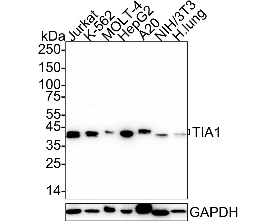
Western blot analysis of TIA1 on different lysates with Rabbit anti-TIA1 antibody at 1/2,000 dilution. Lane 1: Jurkat cell lysate (15 µg/Lane), Lane 2: K-562 cell lysate (15 µg/Lane), Lane 3: MOLT-4 cell lysate (15 µg/Lane), Lane 4: HepG2 cell lysate (15 µg/Lane), Lane 5: A20 cell lysate (15 µg/Lane), Lane 6: NIH/3T3 cell lysate (15 µg/Lane), Lane 7: Human lung tissue lysate (30 µg/Lane), Exposure time: 2 minutes 6 seconds; 4-20% SDS-PAGE gel. Proteins were transferred to a PVDF membrane and blocked with 5% NFDM/TBST for 1 hour at room temperature. The primary antibody at 1/2,000 dilution was used in 5% NFDM/TBST at 4℃ overnight. Goat Anti-Rabbit IgG - HRP Secondary Antibody at 1/50,000 dilution was used for 1 hour at room temperature.IHC
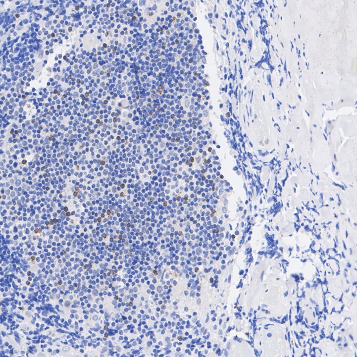
Immunohistochemical analysis of paraffin-embedded human tonsil tissue with Rabbit anti-TIA1 antibody at 1/200 dilution. The section was pre-treated using heat mediated antigen retrieval with sodium citrate buffer (pH 6.0) (high pressure) for 2 minutes. The tissues were blocked in 1% BSA for 20 minutes at room temperature, washed with ddH2O and PBS, and then probed with the primary antibody at 1/200 dilution for 1 hour at room temperature. The detection was performed using an HRP conjugated compact polymer system. DAB was used as the chromogen. Tissues were counterstained with hematoxylin and mounted with DPX.IHC
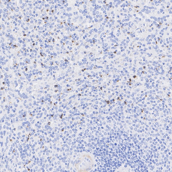
Immunohistochemical analysis of paraffin-embedded human spleen tissue with Rabbit anti-TIA1 antibody at 1/1,000 dilution. The section was pre-treated using heat mediated antigen retrieval with sodium citrate buffer (pH 6.0) (high pressure) for 2 minutes. The tissues were blocked in 1% BSA for 20 minutes at room temperature, washed with ddH2O and PBS, and then probed with the primary antibody at 1/1,000 dilution for 1 hour at room temperature. The detection was performed using an HRP conjugated compact polymer system. DAB was used as the chromogen. Tissues were counterstained with hematoxylin and mounted with DPX.IF-P
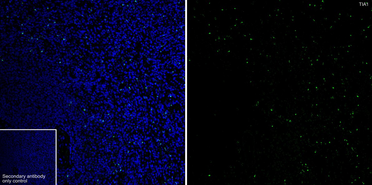
Immunofluorescence analysis of paraffin-embedded human spleen tissue labeling TIA1 with Rabbit anti-TIA1 antibody at 1/50 dilution. The section was pre-treated using heat mediated antigen retrieval with Tris-EDTA buffer (pH 9.0) for 20 minutes. The tissues were blocked in 10% negative goat serum for 1 hour at room temperature, washed with PBS, and then probed with the primary antibody (green) at 1/50 dilution overnight at 4 ℃, washed with PBS. Goat Anti-Rabbit IgG H&L (488) was used as the secondary antibody at 1/1,000 dilution. Nuclei were counterstained with DAPI (blue).| Product Name | TIA1 Recombinant Rabbit Monoclonal Antibody |
|---|---|
| Antibody Type | Primary Antibodies |
| Product description | FAS, also referred to as CD95 or APO-1, is a type I transmembrane protein that plays a central role mediating viral immunity. TIA-1 and TIAR are two closely related proteins that possess three RRMs (RNA recognition motifs), designated RRM 1, 2 and 3. Although both TIA-1 and TIAR are thought to function as mediators of apoptotic cell death, their specific roles in such pathways are unknown. Unlike TIA-1, which is found in the granules of cytotoxic lymphocytes, TIAR expression is limited to the nucleus and found in a much broader range of cells including, but not limited to, cells of hematopoietic origin. TIAR is translocated to the cytoplasm shortly after FAS ligation and this event immediately proceeds the onset of DNA fragmentation. A novel serine/threonine kinase that is activated as a result of FAS ligation, designated FAST (FAS-activated serine/threonine), shows kinase specificity towards both TIA-1 and TIAR. In unstimulated Jurkat cells, FAST resides in the cytoplasm as a highly phosphorylated protein and is quickly dephosphorylated and activated in response to stimulated FAS. |
| Immunogen | Synthetic peptide within Human TIA1 aa 353-386 / 386. |
| Clonality | Monoclonal |
|---|---|
| Isotype | IgG |
| Host Species | Recombinant rabbit |
| Tested Applications | IF-PIHCWB |
| WB:1:500-1:2000 IHC:1:200-1:1000 IF-P:1:50 |
|
| Species Reactivity | HumanMouse |
| Concentration | 1mg/ml |
| Purification | Protein A |
| Alternative Names | Cytotoxic granule associated RNA binding protein 1 antibody Cytotoxic granule associated RNA binding protein antibody mTIA-1 antibody Nucleolysin TIA 1 isoform p40 antibody Nucleolysin TIA-1 isoform p40 antibody Nucleolysin TIA1 isoform p40 antibody p40 TIA 1 antibody p40-TIA-1 (containing p15-TIA-1) antibody p40-TIA-1 antibody RNA binding protein TIA 1 antibody RNA binding protein TIA1 antibody RNA-binding protein TIA-1 antibody T-cell-restricted intracellular antigen-1 antibody TIA 1 antibody TIA 1 cytotoxic granule associated RNA binding protein antibody Tia antibody TIA-1 antibody TIA1 antibody TIA1 cytotoxic granule associated RNA binding protein antibody TIA1 cytotoxic granule associated RNA binding protein like 1 antibody TIA1 protein antibody TIA1_HUMAN antibody TIAL1 antibody TIAR antibody WDM antibody |
|---|---|
| Molecular Weight(MW) | 43 kDa |
| Cellular Localization | Nucleus, Cytoplasm, Stress granule. |
WB
Western blot analysis of TIA1 on different lysates with Rabbit anti-TIA1 antibody at 1/2,000 dilution. Lane 1: Jurkat cell lysate (15 µg/Lane), Lane 2: K-562 cell lysate (15 µg/Lane), Lane 3: MOLT-4 cell lysate (15 µg/Lane), Lane 4: HepG2 cell lysate (15 µg/Lane), Lane 5: A20 cell lysate (15 µg/Lane), Lane 6: NIH/3T3 cell lysate (15 µg/Lane), Lane 7: Human lung tissue lysate (30 µg/Lane), Exposure time: 2 minutes 6 seconds; 4-20% SDS-PAGE gel. Proteins were transferred to a PVDF membrane and blocked with 5% NFDM/TBST for 1 hour at room temperature. The primary antibody at 1/2,000 dilution was used in 5% NFDM/TBST at 4℃ overnight. Goat Anti-Rabbit IgG - HRP Secondary Antibody at 1/50,000 dilution was used for 1 hour at room temperature.
IHC
Immunohistochemical analysis of paraffin-embedded human tonsil tissue with Rabbit anti-TIA1 antibody at 1/200 dilution. The section was pre-treated using heat mediated antigen retrieval with sodium citrate buffer (pH 6.0) (high pressure) for 2 minutes. The tissues were blocked in 1% BSA for 20 minutes at room temperature, washed with ddH2O and PBS, and then probed with the primary antibody at 1/200 dilution for 1 hour at room temperature. The detection was performed using an HRP conjugated compact polymer system. DAB was used as the chromogen. Tissues were counterstained with hematoxylin and mounted with DPX.
IHC
Immunohistochemical analysis of paraffin-embedded human spleen tissue with Rabbit anti-TIA1 antibody at 1/1,000 dilution. The section was pre-treated using heat mediated antigen retrieval with sodium citrate buffer (pH 6.0) (high pressure) for 2 minutes. The tissues were blocked in 1% BSA for 20 minutes at room temperature, washed with ddH2O and PBS, and then probed with the primary antibody at 1/1,000 dilution for 1 hour at room temperature. The detection was performed using an HRP conjugated compact polymer system. DAB was used as the chromogen. Tissues were counterstained with hematoxylin and mounted with DPX.
IF-P
Immunofluorescence analysis of paraffin-embedded human spleen tissue labeling TIA1 with Rabbit anti-TIA1 antibody at 1/50 dilution. The section was pre-treated using heat mediated antigen retrieval with Tris-EDTA buffer (pH 9.0) for 20 minutes. The tissues were blocked in 10% negative goat serum for 1 hour at room temperature, washed with PBS, and then probed with the primary antibody (green) at 1/50 dilution overnight at 4 ℃, washed with PBS. Goat Anti-Rabbit IgG H&L (488) was used as the secondary antibody at 1/1,000 dilution. Nuclei were counterstained with DAPI (blue).| Application Notes | WB:1:500-1:2000 IHC:1:200-1:1000 IF-P:1:50 |
|---|
| Form | Liquid |
|---|---|
| Storage Instructions | Store at +4℃ after thawing. Aliquot store at -20℃ or -80℃. Avoid repeated freeze / thaw cycles. |
| Storage Buffer | 1*TBS (pH7.4), 1%BSA, 40%Glycerol. Preservative: 0.05% Sodium Azide. |


