WB
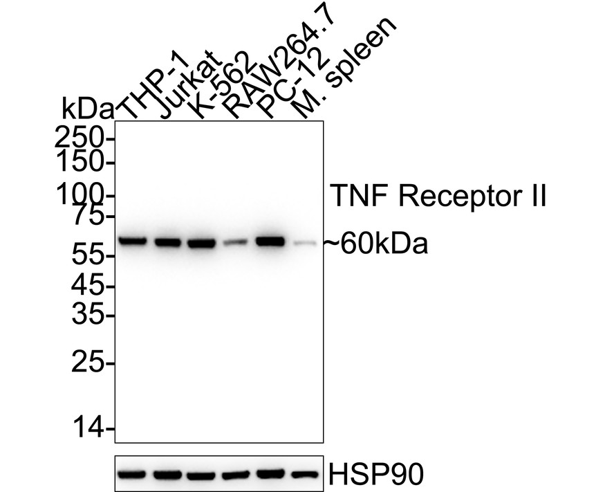
Western blot analysis of TNF Receptor II on different lysates with Rabbit anti-TNF Receptor II antibody at 1/2,000 dilution. Lane 1: THP-1 cell lysate (20 µg/Lane), Lane 2: Jurkat cell lysate (20 µg/Lane), Lane 3: K-562 cell lysate (20 µg/Lane), Lane 4: RAW264.7 cell lysate (20 µg/Lane), Lane 5: PC-12 cell lysate (20 µg/Lane), Lane 6: Mouse spleen tissue lysate (40 µg/Lane), Exposure time: 10 seconds; 4-20% SDS-PAGE gel. Proteins were transferred to a PVDF membrane and blocked with 5% NFDM/TBST for 1 hour at room temperature. The primary antibody at 1/2,000 dilution was used in 5% NFDM/TBST at 4℃ overnight. Goat Anti-Rabbit IgG - HRP Secondary Antibody at 1/50,000 dilution was used for 1 hour at room temperature.IHC
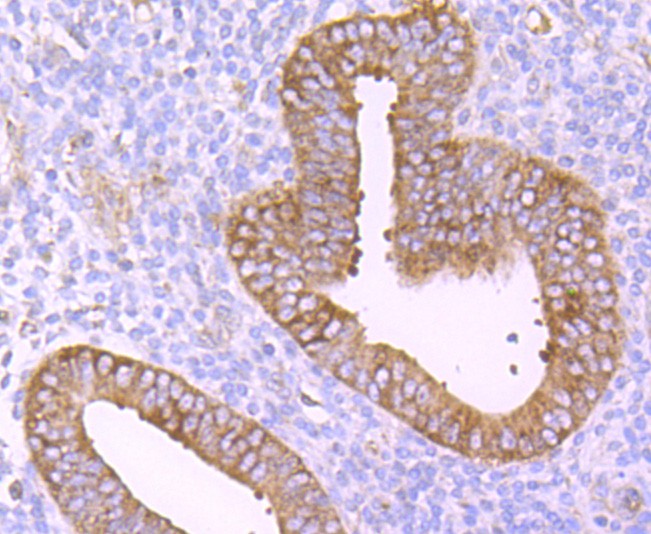
Immunohistochemical analysis of paraffin-embedded human uterus tissue using anti-TNF Receptor II antibody. The section was pre-treated using heat mediated antigen retrieval with Tris-EDTA buffer (pH 8.0-8.4) for 20 minutes.The tissues were blocked in 5% BSA for 30 minutes at room temperature, washed with ddH2O and PBS, and then probed with the primary antibody (1/50) for 30 minutes at room temperature. The detection was performed using an HRP conjugated compact polymer system. DAB was used as the chromogen. Tissues were counterstained with hematoxylin and mounted with DPX.ICC/IF
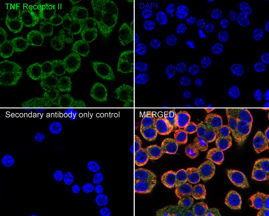
Immunocytochemistry analysis of RAW264.7 cells labeling TNF Receptor II with Rabbit anti-TNF Receptor II antibody at 1/100 dilution. Cells were fixed in 4% paraformaldehyde for 20 minutes at room temperature, permeabilized with 0.1% Triton X-100 in PBS for 5 minutes at room temperature, then blocked with 1% BSA in 10% negative goat serum for 1 hour at room temperature. Cells were then incubated with Rabbit anti-TNF Receptor II antibody at 1/100 dilution in 1% BSA in PBST overnight at 4 ℃. Goat Anti-Rabbit IgG H&L (488) was used as the secondary antibody at 1/1,000 dilution. PBS instead of the primary antibody was used as the secondary antibody only control. Nuclear DNA was labelled in blue with DAPI. Beta tubulin (red) was stained at 1/100 dilution overnight at +4℃. Goat Anti-Mouse IgG H&L (594) was used as the secondary antibody at 1/1,000 dilution.IF-F
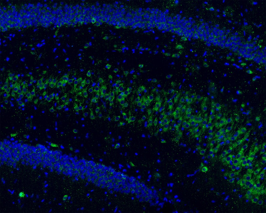
Immunofluorescence analysis of frozen mouse hippocampus tissue labeling TNF Receptor II with Rabbit anti-TNF Receptor II antibody. The tissues were blocked in 3% BSA for 30 minutes at room temperature, washed with PBS, and then probed with the primary antibody (green) at 1/100 dilution overnight at 4℃, washed with PBS. Goat Anti-Rabbit IgG H&L (Alexa Fluor® 488) was used as the secondary antibody at 1/200 dilution. Nuclei were counterstained with DAPI (blue). Image acquisition was performed with KFBIO KF-FL-400 Scanner.IF-F
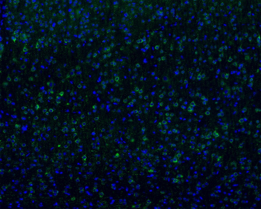
Immunofluorescence analysis of frozen mouse cerebral cortex tissue labeling TNF Receptor II with Rabbit anti-TNF Receptor II antibody. The tissues were blocked in 3% BSA for 30 minutes at room temperature, washed with PBS, and then probed with the primary antibody (green) at 1/100 dilution overnight at 4℃, washed with PBS. Goat Anti-Rabbit IgG H&L (Alexa Fluor® 488) was used as the secondary antibody at 1/200 dilution. Nuclei were counterstained with DAPI (blue). Image acquisition was performed with KFBIO KF-FL-400 Scanner.FC
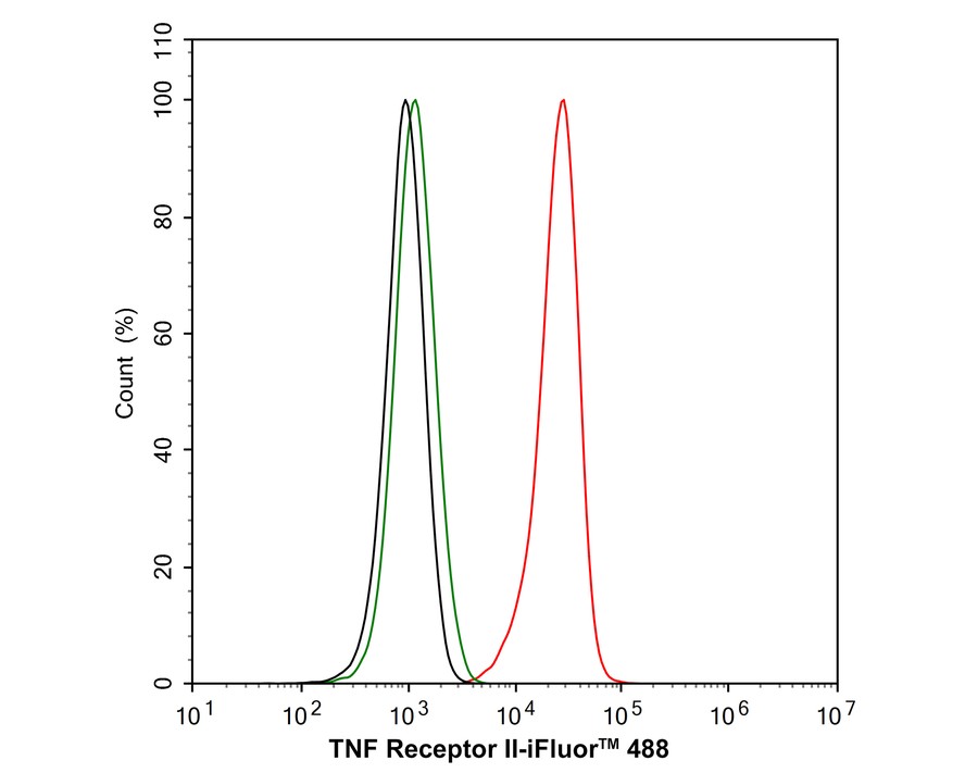
Flow cytometric analysis of RAW264.7 cells labeling TNF Receptor II. Cells were fixed and permeabilized. Then stained with the primary antibody (1μg/mL) (red) compared with Rabbit IgG Isotype Control (green). After incubation of the primary antibody at +4℃ for an hour, the cells were stained with a 488 conjugate-Goat anti-Rabbit IgG Secondary antibody at 1/1,000 dilution for 30 minutes at +4℃. Unlabelled sample was used as a control (cells without incubation with primary antibody; black).IP
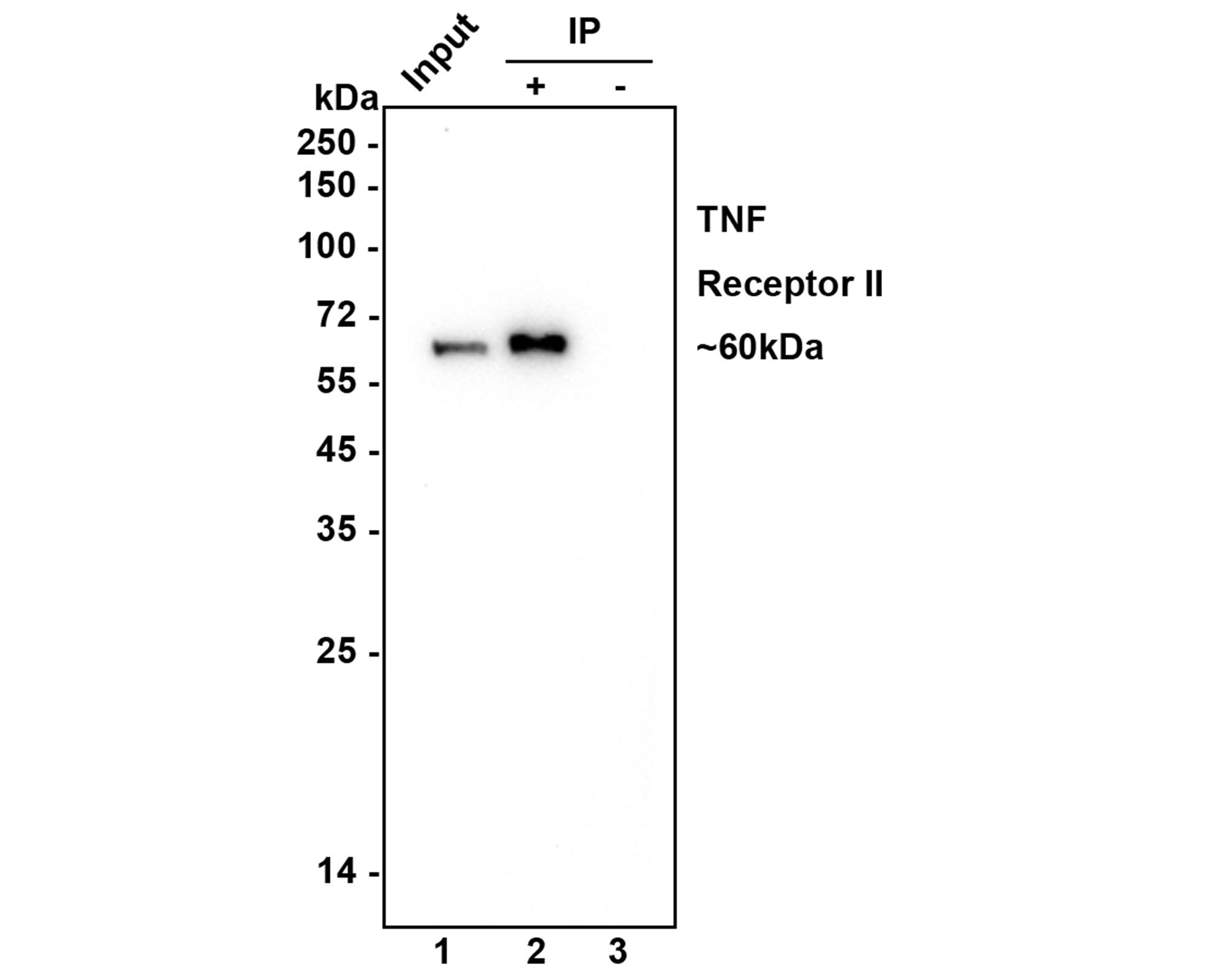
TNF Receptor II was immunoprecipitated in 0.2mg Jurkat cell lysate with Rabbit anti-TNF Receptor II antibody at 2 µg/10 µl beads. Western blot was performed from the immunoprecipitate using Rabbit anti-TNF Receptor II antibody at 1/1,000 dilution. Anti-Rabbit IgG for IP Nano-secondary antibody at 1/5,000 dilution was used for 1 hour at room temperature. Lane 1: Jurkat cell lysate (input), Lane 2: Rabbit anti-TNF Receptor II antibody IP in Jurkat cell lysate, Lane 3: Rabbit IgG instead of Rabbit anti-TNF Receptor II antibody in Jurkat cell lysate. Blocking/Dilution buffer: 5% NFDM/TBST, Exposure time: 3 mintues.| Product Name | TNF Receptor II Recombinant Rabbit Monoclonal Antibody |
|---|---|
| Antibody Type | Primary Antibodies |
| Product description | Tumor necrosis factor (TNF) is a pleiotropic cytokine whose function is mediated through two distinct cell surface receptors. These receptors, designated TNF-R1 and TNF-R2, are expressed on most cell types. The majority of TNF functions are primarily mediated through TNF-R1, while signaling through TNF-R2 occurs less extensively and is confined to cells of the immune system. Both of these proteins belong to the growing TNF and nerve growth factor (NGF) receptor superfamily, which includes FAS, CD30, CD27 and CD40. The members of this superfamily are type I membrane proteins that share sequence homology confined to the extracellular region. TNF-R1 shares a motif termed the "death domain" with FAS and three structurally unrelated signaling proteins, TRADD, FADD and RIP (1,3-8). This death domain is required for transduction of the apoptotic signal. |
| Immunogen | Synthetic peptide within Human TNF Receptor II aa 426-461 / 461. |
| Clonality | Monoclonal |
|---|---|
| Isotype | IgG |
| Host Species | Recombinant rabbit |
| Tested Applications | FCICC/IFIF-FIHCIPWB |
| WB:1:2000 IHC:1:50-1:200 ICC/IF:1:50-1:200 IF-F:1:200 IF-F:1:200 FC:1:1000 IP:Use at an assay dependent concentration. |
|
| Species Reactivity | HumanMouseRat |
| Concentration | 1mg/ml |
| Purification | Protein A |
| Alternative Names | CD120b antibody p75 antibody p75 TNF receptor antibody p75TNFR antibody p80 TNF alpha receptor antibody p80 TNF-alpha receptor antibody Soluble TNFR1B variant 1 antibody TBP-2 antibody TBPII antibody TNF R II antibody TNF R2 antibody TNF R75 antibody TNF-R2 antibody TNF-RII antibody TNFBR antibody TNFR-II antibody TNFR1B antibody TNFR2 antibody TNFR80 antibody TNFRII antibody Tnfrsf1b antibody TNR1B_HUMAN antibody Tumor necrosis factor beta receptor antibody Tumor necrosis factor receptor 2 antibody Tumor necrosis factor receptor superfamily member 1B antibody Tumor necrosis factor receptor type II antibody Tumor necrosis factor-binding protein 2 antibody |
|---|---|
| Molecular Weight(MW) | 48 kDa(Observed band size: 60 kDa) |
| Cellular Localization | Cell membrane, Membrane, Secreted. |
WB
Western blot analysis of TNF Receptor II on different lysates with Rabbit anti-TNF Receptor II antibody at 1/2,000 dilution. Lane 1: THP-1 cell lysate (20 µg/Lane), Lane 2: Jurkat cell lysate (20 µg/Lane), Lane 3: K-562 cell lysate (20 µg/Lane), Lane 4: RAW264.7 cell lysate (20 µg/Lane), Lane 5: PC-12 cell lysate (20 µg/Lane), Lane 6: Mouse spleen tissue lysate (40 µg/Lane), Exposure time: 10 seconds; 4-20% SDS-PAGE gel. Proteins were transferred to a PVDF membrane and blocked with 5% NFDM/TBST for 1 hour at room temperature. The primary antibody at 1/2,000 dilution was used in 5% NFDM/TBST at 4℃ overnight. Goat Anti-Rabbit IgG - HRP Secondary Antibody at 1/50,000 dilution was used for 1 hour at room temperature.
IHC
Immunohistochemical analysis of paraffin-embedded human uterus tissue using anti-TNF Receptor II antibody. The section was pre-treated using heat mediated antigen retrieval with Tris-EDTA buffer (pH 8.0-8.4) for 20 minutes.The tissues were blocked in 5% BSA for 30 minutes at room temperature, washed with ddH2O and PBS, and then probed with the primary antibody (1/50) for 30 minutes at room temperature. The detection was performed using an HRP conjugated compact polymer system. DAB was used as the chromogen. Tissues were counterstained with hematoxylin and mounted with DPX.
ICC/IF
Immunocytochemistry analysis of RAW264.7 cells labeling TNF Receptor II with Rabbit anti-TNF Receptor II antibody at 1/100 dilution. Cells were fixed in 4% paraformaldehyde for 20 minutes at room temperature, permeabilized with 0.1% Triton X-100 in PBS for 5 minutes at room temperature, then blocked with 1% BSA in 10% negative goat serum for 1 hour at room temperature. Cells were then incubated with Rabbit anti-TNF Receptor II antibody at 1/100 dilution in 1% BSA in PBST overnight at 4 ℃. Goat Anti-Rabbit IgG H&L (488) was used as the secondary antibody at 1/1,000 dilution. PBS instead of the primary antibody was used as the secondary antibody only control. Nuclear DNA was labelled in blue with DAPI. Beta tubulin (red) was stained at 1/100 dilution overnight at +4℃. Goat Anti-Mouse IgG H&L (594) was used as the secondary antibody at 1/1,000 dilution.
IF-F
Immunofluorescence analysis of frozen mouse hippocampus tissue labeling TNF Receptor II with Rabbit anti-TNF Receptor II antibody. The tissues were blocked in 3% BSA for 30 minutes at room temperature, washed with PBS, and then probed with the primary antibody (green) at 1/100 dilution overnight at 4℃, washed with PBS. Goat Anti-Rabbit IgG H&L (Alexa Fluor® 488) was used as the secondary antibody at 1/200 dilution. Nuclei were counterstained with DAPI (blue). Image acquisition was performed with KFBIO KF-FL-400 Scanner.
IF-F
Immunofluorescence analysis of frozen mouse cerebral cortex tissue labeling TNF Receptor II with Rabbit anti-TNF Receptor II antibody. The tissues were blocked in 3% BSA for 30 minutes at room temperature, washed with PBS, and then probed with the primary antibody (green) at 1/100 dilution overnight at 4℃, washed with PBS. Goat Anti-Rabbit IgG H&L (Alexa Fluor® 488) was used as the secondary antibody at 1/200 dilution. Nuclei were counterstained with DAPI (blue). Image acquisition was performed with KFBIO KF-FL-400 Scanner.
FC
Flow cytometric analysis of RAW264.7 cells labeling TNF Receptor II. Cells were fixed and permeabilized. Then stained with the primary antibody (1μg/mL) (red) compared with Rabbit IgG Isotype Control (green). After incubation of the primary antibody at +4℃ for an hour, the cells were stained with a 488 conjugate-Goat anti-Rabbit IgG Secondary antibody at 1/1,000 dilution for 30 minutes at +4℃. Unlabelled sample was used as a control (cells without incubation with primary antibody; black).
IP
TNF Receptor II was immunoprecipitated in 0.2mg Jurkat cell lysate with Rabbit anti-TNF Receptor II antibody at 2 µg/10 µl beads. Western blot was performed from the immunoprecipitate using Rabbit anti-TNF Receptor II antibody at 1/1,000 dilution. Anti-Rabbit IgG for IP Nano-secondary antibody at 1/5,000 dilution was used for 1 hour at room temperature. Lane 1: Jurkat cell lysate (input), Lane 2: Rabbit anti-TNF Receptor II antibody IP in Jurkat cell lysate, Lane 3: Rabbit IgG instead of Rabbit anti-TNF Receptor II antibody in Jurkat cell lysate. Blocking/Dilution buffer: 5% NFDM/TBST, Exposure time: 3 mintues.| Application Notes | WB:1:2000 IHC:1:50-1:200 ICC/IF:1:50-1:200 IF-F:1:200 IF-F:1:200 FC:1:1000 IP:Use at an assay dependent concentration. |
|---|
| Form | Liquid |
|---|---|
| Storage Instructions | Store at +4℃ after thawing. Aliquot store at -20℃ or -80℃. Avoid repeated freeze / thaw cycles. |
| Storage Buffer | 1*TBS (pH7.4), 1%BSA, 40%Glycerol. Preservative: 0.05% Sodium Azide. |


