Application
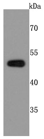
Fig1: Western blot analysis of PABPN1 on MCF-7 cells lysates using anti-PABPN1 antibody at 1/500 dilution.Application
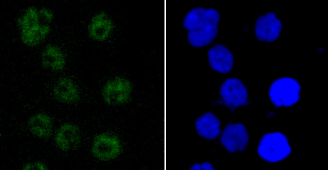
Fig2: ICC staining PABPN1 in AGS cells (green). The nuclear counter stain is DAPI (blue). Cells were fixed in paraformaldehyde, permeabilised with 0.25% Triton X100/PBS.Application
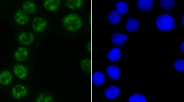
Fig3: ICC staining PABPN1 in SW480 cells (green). The nuclear counter stain is DAPI (blue). Cells were fixed in paraformaldehyde, permeabilised with 0.25% Triton X100/PBS.Application
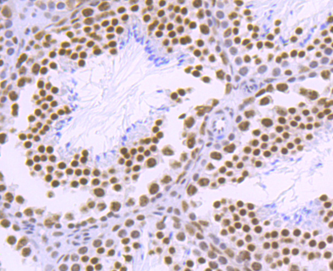
Fig4: Immunohistochemical analysis of paraffin-embedded mouse testis tissue using anti-PABPN1 antibody. Counter stained with hematoxylin.Application
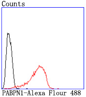
Fig5: Flow cytometric analysis of Hela cells with PABPN1 antibody at 1/50 dilution (red) compared with an unlabelled control (cells without incubation with primary antibody; black). Alexa Fluor 488-conjugated goat anti rabbit IgG was used as the secondary antibody.| Product Name | PABPN1 [JM11-28] |
|---|---|
| Antibody Type | Primary Antibodies |
| Antigen Alias | Nuclear poly(A)-binding protein 1 antibody OPMD antibody PAB2 antibody PABII antibody PABP 2 antibody pABP-2 antibody PABP2 antibody PABP2_HUMAN antibody PABPII antibody Pabpn1 antibody poly(A) binding protein nuclear 1 antibody Poly(A)-binding protein 2 antibody Poly(A)-binding protein II antibody PolyA binding protein II antibody Polyadenylate-binding nuclear protein 1 antibody Polyadenylate-binding protein 2 antibody |
| Product description | Involved in the 3'-end formation of mRNA precursors (pre-mRNA) by the addition of a poly(A) tail of 200-250 nt to the upstream cleavage product. Stimulates poly(A) polymerase (PAPOLA) conferring processivity on the poly(A) tail elongation reaction and controls also the poly(A) tail length. Increases the affinity of poly(A) polymerase for RNA. Is also present at various stages of mRNA metabolism including nucleocytoplasmic trafficking and nonsense-mediated decay (NMD) of mRNA. Cooperates with SKIP to synergistically activate E-box-mediated transcription through MYOD1 and may regulate the expression of muscle-specific genes. Binds to poly(A) and to poly(G) with high affinity. May protect the poly(A) tail from degradation. |
| Immunogen | recombinant protein |
| Clonality | Monoclonal |
|---|---|
| Isotype | IgG |
| Host Species | Recombinant rabbit |
| Tested Applications | WBICC/IFIHCFCIP |
| WB:1:500-1:2,000 ICC:1:50-1:200 IHC:1:50-1:200 FC:1:50-1:100 IP:1:10-1:50 |
|
| Species Reactivity | HumanMouse |
| Concentration | 1mg/ml |
| Alternative Names | Nuclear poly(A)-binding protein 1 antibody OPMD antibody PAB2 antibody PABII antibody PABP 2 antibody pABP-2 antibody PABP2 antibody PABP2_HUMAN antibody PABPII antibody Pabpn1 antibody poly(A) binding protein nuclear 1 antibody Poly(A)-binding protein 2 antibody Poly(A)-binding protein II antibody PolyA binding protein II antibody Polyadenylate-binding nuclear protein 1 antibody Polyadenylate-binding protein 2 antibody |
|---|---|
| Molecular Weight(MW) | 49 kDa |
| Cellular Localization | Nucleus. Cytoplasm. |
Application
Fig1: Western blot analysis of PABPN1 on MCF-7 cells lysates using anti-PABPN1 antibody at 1/500 dilution.
Application
Fig2: ICC staining PABPN1 in AGS cells (green). The nuclear counter stain is DAPI (blue). Cells were fixed in paraformaldehyde, permeabilised with 0.25% Triton X100/PBS.
Application
Fig3: ICC staining PABPN1 in SW480 cells (green). The nuclear counter stain is DAPI (blue). Cells were fixed in paraformaldehyde, permeabilised with 0.25% Triton X100/PBS.
Application
Fig4: Immunohistochemical analysis of paraffin-embedded mouse testis tissue using anti-PABPN1 antibody. Counter stained with hematoxylin.
Application
Fig5: Flow cytometric analysis of Hela cells with PABPN1 antibody at 1/50 dilution (red) compared with an unlabelled control (cells without incubation with primary antibody; black). Alexa Fluor 488-conjugated goat anti rabbit IgG was used as the secondary antibody.| Positive Control | MCF-7, AGS, SW480, mouse testis tissue. |
|---|---|
| Application Notes | WB:1:500-1:2,000 ICC:1:50-1:200 IHC:1:50-1:200 FC:1:50-1:100 IP:1:10-1:50 |
| Form | Liquid |
|---|---|
| Storage Instructions | Store at +4℃ after thawing. Aliquot store at -20℃ or -80℃. Avoid repeated freeze / thaw cycles. |
| Storage Buffer | 1*TBS (pH7.4), 1%BSA, 40%Glycerol. Preservative: 0.05% Sodium Azide. |


