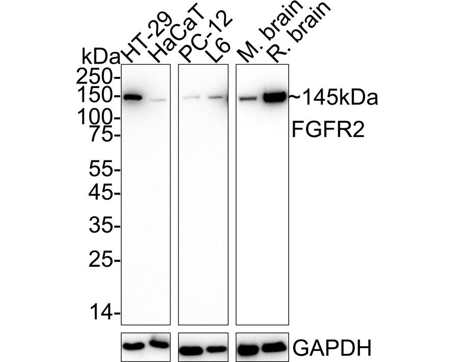
WB
Western blot analysis of FGFR2 on different lysates with Rabbit anti-FGFR2 antibody at 1/1,000 dilution. Lane 1: HT-29 cell lysate (20 µg/Lane), Lane 2: HaCaT cell lysate (20 µg/Lane), Lane 3: PC-12 cell lysate (20 µg/Lane), Lane 4: L6 cell lysate (20 µg/Lane), Lane 5: Mouse brain tissue lysate (40 µg/Lane), Lane 6: Rat brain tissue lysate (40 µg/Lane), Exposure time: Lane 1-4: 1 minute 18 seconds; Lane 5-6: 6 seconds; 4-20% SDS-PAGE gel. Proteins were transferred to a PVDF membrane and blocked with 5% NFDM/TBST for 1 hour at room temperature. The primary antibody at 1/1,000 dilution was used in 5% NFDM/TBST at 4℃ overnight. Goat Anti-Rabbit IgG - HRP Secondary Antibody at 1/50,000 dilution was used for 1 hour at room temperature.| Product Name | FGFR2 Recombinant Rabbit Monoclonal Antibody |
|---|---|
| Antibody Type | Primary Antibodies |
| Product description | Acidic and basic fibroblast growth factors (FGFs) are members of a family of multifunctional polypeptide growth factors that stimulate proliferation of cells of mesenchymal, epithelial and neuroectodermal origin. Like other growth factors, FGFs act by binding and activating specific cell surface receptors. These include the Flg receptor or FGFR-1, the Bek receptor (or FGFR-2), FGFR-3, FGFR-4, FGFR-5 and FGFR-6. These receptors usually contain an extracellular ligand-binding region containing three immunoglobulin-like domains, a transmembrane domain and a cytoplasmic tyrosine kinase domain. The gene encoding human Bek (also designated K-sam) maps to chromosome 10q26.13 and is alternatively spliced to produce several isoforms. Heterogeneous mutations in Bek are associated with a range of craniosynostosis syndromes including Pfeiffer syndrome, Crouzon syndrome, Jackson-Weiss syndrome and Apert syndrome.. |
| Immunogen | Synthetic peptide within Human FGFR2 aa 30-70 / 821. |
| Clonality | Monoclonal |
|---|---|
| Isotype | IgG |
| Host Species | Recombinant rabbit |
| Tested Applications | WB |
| WB:1:1000-1:2000 |
|
| Species Reactivity | HumanMouseRat |
| Concentration | 1mg/ml |
| Purification | Protein A |
| Alternative Names | bacteria-expressed kinase antibody
BBDS antibody
BEK antibody
BEK fibroblast growth factor receptor antibody
BFR1 antibody
CD332 antibody
CD332 antigen antibody
CEK3 antibody
CFD1 antibody
Craniofacial dysostosis 1 antibody
ECT1 antibody
FGF receptor antibody
FGFR 2 antibody
FGFR-2 antibody
Fgfr2 antibody
FGFR2_HUMAN antibody
Fibroblast growth factor receptor 2 antibody
Hydroxyaryl protein kinase antibody
Jackson Weiss syndrome antibody
JWS antibody
K SAM antibody
K-sam antibody
Keratinocyte growth factor receptor 2 antibody
Keratinocyte growth factor receptor antibody
KGFR antibody
KSAM antibody
protein tyrosine kinase receptor like 14 antibody soluble FGFR4 variant 4 antibody TK14 antibody TK25 antibody |
|---|---|
| Molecular Weight(MW) | 145 kDa |
| Cellular Localization | Cell membrane, Golgi apparatus, Cytoplasmic vesicle. |
WB
Western blot analysis of FGFR2 on different lysates with Rabbit anti-FGFR2 antibody at 1/1,000 dilution. Lane 1: HT-29 cell lysate (20 µg/Lane), Lane 2: HaCaT cell lysate (20 µg/Lane), Lane 3: PC-12 cell lysate (20 µg/Lane), Lane 4: L6 cell lysate (20 µg/Lane), Lane 5: Mouse brain tissue lysate (40 µg/Lane), Lane 6: Rat brain tissue lysate (40 µg/Lane), Exposure time: Lane 1-4: 1 minute 18 seconds; Lane 5-6: 6 seconds; 4-20% SDS-PAGE gel. Proteins were transferred to a PVDF membrane and blocked with 5% NFDM/TBST for 1 hour at room temperature. The primary antibody at 1/1,000 dilution was used in 5% NFDM/TBST at 4℃ overnight. Goat Anti-Rabbit IgG - HRP Secondary Antibody at 1/50,000 dilution was used for 1 hour at room temperature.| Application Notes | WB:1:1000-1:2000 |
|---|
| Form | Liquid |
|---|---|
| Storage Instructions | Store at +4℃ after thawing. Aliquot store at -20℃ or -80℃. Avoid repeated freeze / thaw cycles. |
| Storage Buffer | 1*TBS (pH7.4), 1%BSA, 40%Glycerol. Preservative: 0.05% Sodium Azide. |


