Application
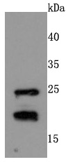
Fig1: Western blot analysis of RAC1/2/3 on MCF-7 cells lysates using anti-RAC1/2/3 antibody at 1/500 dilution.Application
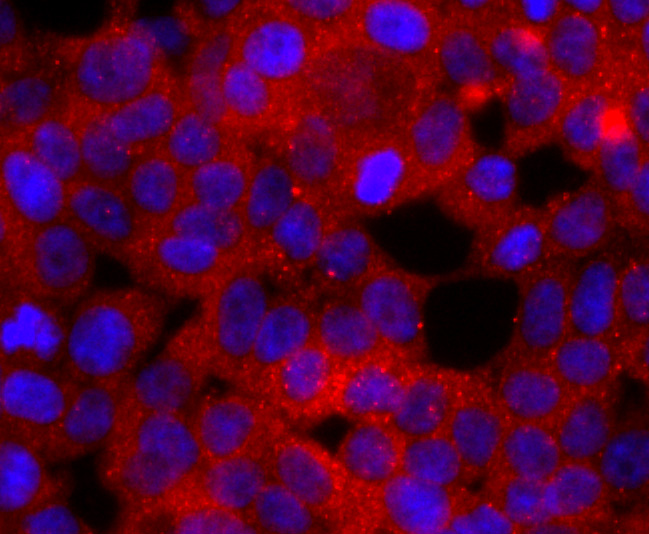
Fig2: ICC staining RAC1/2/3 in 293T cells (red). The nuclear counter stain is DAPI (blue). Cells were fixed in paraformaldehyde, permeabilised with 0.25% Triton X100/PBS.Application
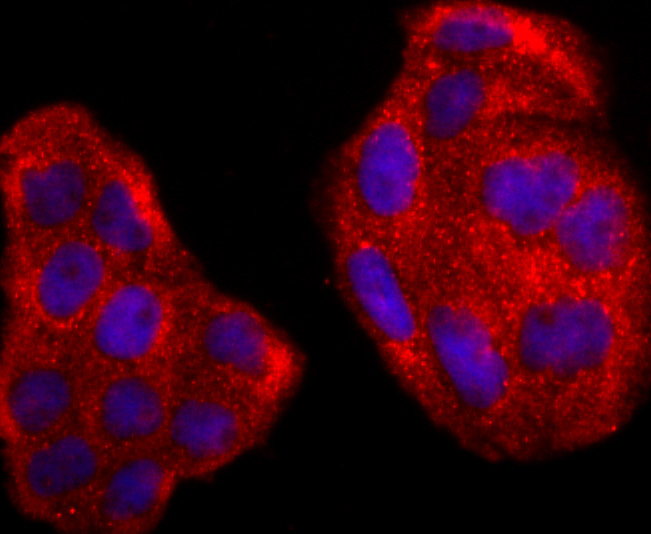
Fig3: ICC staining RAC1/2/3 in Hela cells (red). The nuclear counter stain is DAPI (blue). Cells were fixed in paraformaldehyde, permeabilised with 0.25% Triton X100/PBS.Application
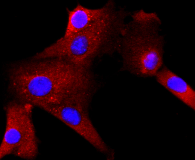
Fig4: ICC staining RAC1/2/3 in NIH-3T3 cells (red). The nuclear counter stain is DAPI (blue). Cells were fixed in paraformaldehyde, permeabilised with 0.25% Triton X100/PBS.Application
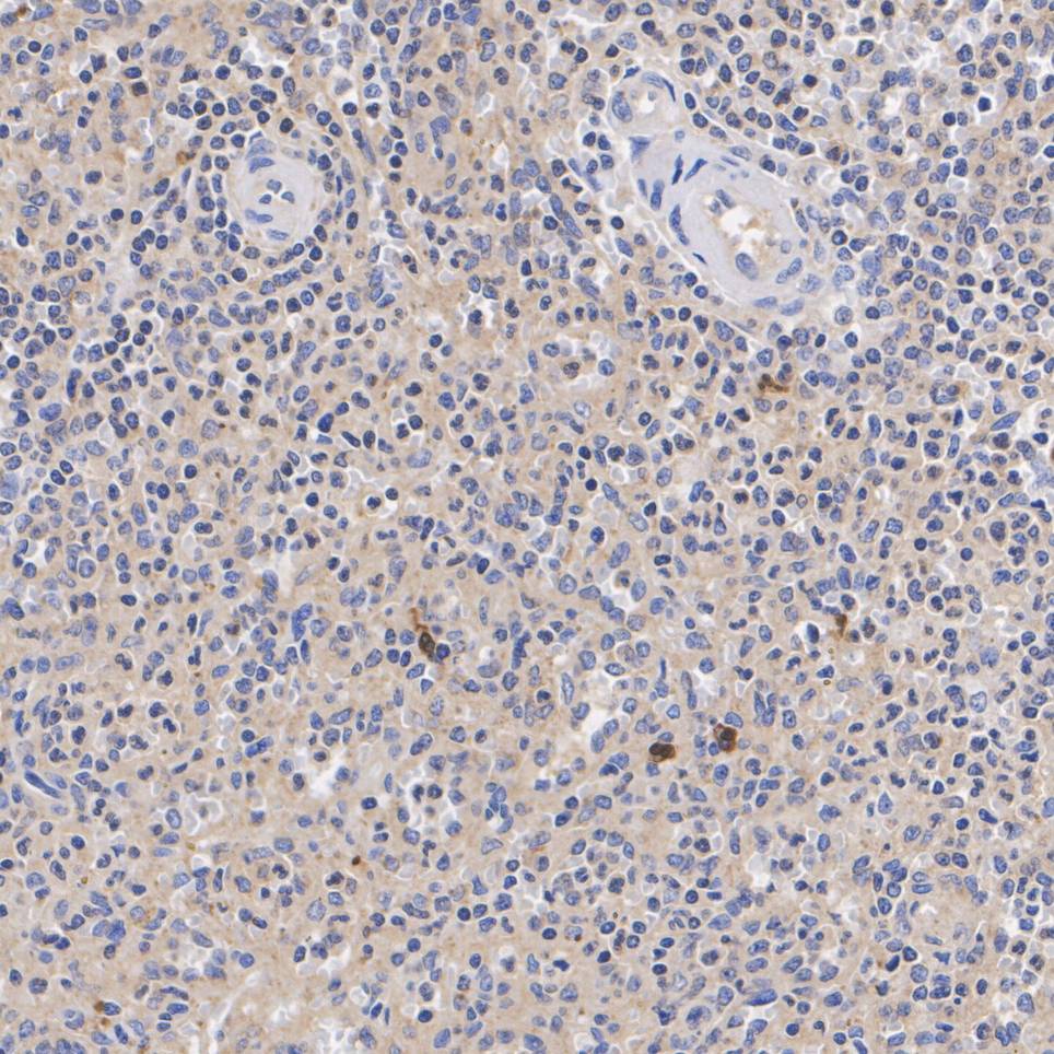
Fig5: Immunohistochemical analysis of paraffin-embedded human spleen tissue with Rabbit anti-Rac1-2-3 antibody at 1/200 dilution. The section was pre-treated using heat mediated antigen retrieval with Tris-EDTA buffer (pH 9.0) for 20 minutes. The tissues were blocked in 1% BSA for 20 minutes at room temperature, washed with ddH2O and PBS, and then probed with the primary antibody at 1/200 dilution for 1 hour at room temperature. The detection was performed using an HRP conjugated compact polymer system. DAB was used as the chromogen. Tissues were counterstained with hematoxylin and mounted with DPX.Application
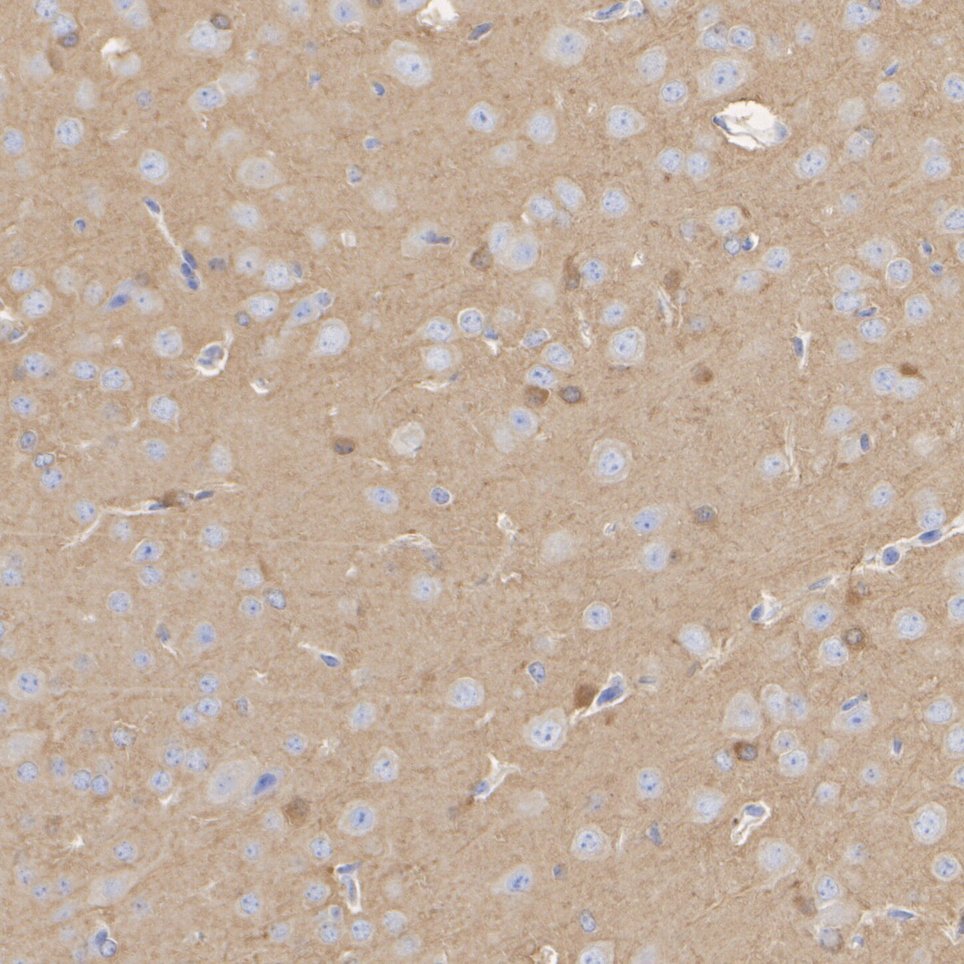
Fig6: Immunohistochemical analysis of paraffin-embedded mouse brain tissue with Rabbit anti-Rac1-2-3 antibody at 1/200 dilution. The section was pre-treated using heat mediated antigen retrieval with Tris-EDTA buffer (pH 9.0) for 20 minutes. The tissues were blocked in 1% BSA for 20 minutes at room temperature, washed with ddH2O and PBS, and then probed with the primary antibody at 1/200 dilution for 1 hour at room temperature. The detection was performed using an HRP conjugated compact polymer system. DAB was used as the chromogen. Tissues were counterstained with hematoxylin and mounted with DPX.Application
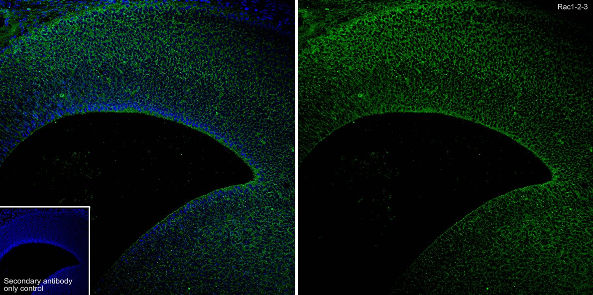
Fig7:Immunofluorescence analysis of paraffin-embedded mouse embryonic brain tissue labeling Rac1-2-3 with Rabbit anti-Rac1-2-3 antibody at 1/200 dilution. The section was pre-treated using heat mediated antigen retrieval with Tris-EDTA buffer (pH 9.0) for 20 minutes. The tissues were blocked in 10% negative goat serum for 1 hour at room temperature, washed with PBS, and then probed with the primary antibody (green) at 1/200 dilution overnight at 4 ℃, washed with PBS. Goat Anti-Rabbit IgG H&L (488) was used as the secondary antibody at 1/1,000 dilution. Nuclei were counterstained with DAPI (blue).| Product Name | Rac1-2-3 Recombinant Rabbit Monoclonal Antibody |
|---|---|
| Antibody Type | Primary Antibodies |
| Product description | Plasma membrane-associated small GTPase which cycles between active GTP-bound and inactive GDP-bound states. In its active state, binds to a variety of effector proteins to regulate cellular responses such as secretory processes, phagocytosis of apoptotic cells, epithelial cell polarization and growth-factor induced formation of membrane ruffles. Rac1 p21/rho GDI heterodimer is the active component of the cytosolic factor sigma 1, which is involved in stimulation of the NADPH oxidase activity in macrophages (By similarity). Essential for the SPATA13-mediated regulation of cell migration and adhesion assembly and disassembly. Isoform B has an accelerated GEF-independent GDP/GTP exchange and an impaired GTP hydrolysis, which is restored partially by GTPase-activating proteins. It is able to bind to the GTPase-binding domain of PAK but not full-length PAK in a GTP-dependent manner, suggesting that the insertion does not completely abolish effector interaction. |
| Immunogen | Recombinant full length protein of Human RAC1 aa 1-192 / 192. |
| Clonality | Monoclonal |
|---|---|
| Isotype | IgG |
| Host Species | Recombinant rabbit |
| Tested Applications | ICC/IFIF-PIHCWB |
| WB:1:500-1:2000 ICC/IF:1:50-1:200 IHC:1:200 IF-P:1:200 |
|
| Species Reactivity | HumanMouseRat |
| Concentration | 1mg/ml |
| Purification | Protein A |
| Alternative Names | Cell migration inducing gene 5 protein antibody
EN 7 antibody
OTTMUSP00000004488 antibody
p21 Rac3 antibody
p21-Rac3 antibody
Rac1B antibody
RAC3 antibody
RAC3_HUMAN antibody
RAS related C3 botulinum substrate 3 antibody
Ras related C3 botulinum toxin substrate 3 (rho family small GTP binding protein Rac3) antibody
Ras related C3 botulinum toxin substrate 3 (rho family small GTP binding protein Rac3) antibody Ras-related C3 botulinum toxin substrate 3 antibody Rho family small GTP binding protein Rac3 antibody RP23-84C12.18 antibody EN-7 antibody EN7 antibody GX antibody HSPC 022 antibody HSPC022 antibody p21 Rac 2 antibody p21 Rac2 antibody p21-Rac2 antibody p21Rac2 antibody RAC 2 antibody Rac2 antibody RAC2_HUMAN antibody Ras related C3 botulinum toxin substrate 2 (rho family small GTP binding protein Rac2 antibody Ras related C3 botulinum toxin substrate 2 antibody Ras related C3 botulinum toxin substrate 3 (rho family small GTP binding protein Rac2) antibody Ras related C3 botulinum toxin substrate 3 antibody Ras-related C3 botulinum toxin substrate 2 antibody Rho family small GTP binding protein Rac 2 antibody Rho family small GTP binding protein Rac2 antibody Small G protein antibody Cell migration-inducing gene 5 protein antibody MGC111543 antibody MIG5 antibody Migration inducing gene 5 antibody Migration inducing protein 5 antibody p21 Rac1 antibody p21-Rac1 antibody Rac 1 antibody RAC1 antibody RAC1_HUMAN antibody Ras like protein TC25 antibody Ras related C3 botulinum toxin substrate 1 (rho family small GTP binding protein Rac1) antibody Ras-like protein TC25 antibody Ras-related C3 botulinum toxin substrate 1 antibody Rho family small GTP binding protein Rac1 antibody TC 25 antibody TC25 antibody |
|---|---|
| Molecular Weight(MW) | 21/24 kDa |
| Cellular Localization | Cell membrane, Cell projection, Cytoplasm, Cytoskeleton, Membrane. |
Application
Fig1: Western blot analysis of RAC1/2/3 on MCF-7 cells lysates using anti-RAC1/2/3 antibody at 1/500 dilution.
Application
Fig2: ICC staining RAC1/2/3 in 293T cells (red). The nuclear counter stain is DAPI (blue). Cells were fixed in paraformaldehyde, permeabilised with 0.25% Triton X100/PBS.
Application
Fig3: ICC staining RAC1/2/3 in Hela cells (red). The nuclear counter stain is DAPI (blue). Cells were fixed in paraformaldehyde, permeabilised with 0.25% Triton X100/PBS.
Application
Fig4: ICC staining RAC1/2/3 in NIH-3T3 cells (red). The nuclear counter stain is DAPI (blue). Cells were fixed in paraformaldehyde, permeabilised with 0.25% Triton X100/PBS.
Application
Fig5: Immunohistochemical analysis of paraffin-embedded human spleen tissue with Rabbit anti-Rac1-2-3 antibody at 1/200 dilution. The section was pre-treated using heat mediated antigen retrieval with Tris-EDTA buffer (pH 9.0) for 20 minutes. The tissues were blocked in 1% BSA for 20 minutes at room temperature, washed with ddH2O and PBS, and then probed with the primary antibody at 1/200 dilution for 1 hour at room temperature. The detection was performed using an HRP conjugated compact polymer system. DAB was used as the chromogen. Tissues were counterstained with hematoxylin and mounted with DPX.
Application
Fig6: Immunohistochemical analysis of paraffin-embedded mouse brain tissue with Rabbit anti-Rac1-2-3 antibody at 1/200 dilution. The section was pre-treated using heat mediated antigen retrieval with Tris-EDTA buffer (pH 9.0) for 20 minutes. The tissues were blocked in 1% BSA for 20 minutes at room temperature, washed with ddH2O and PBS, and then probed with the primary antibody at 1/200 dilution for 1 hour at room temperature. The detection was performed using an HRP conjugated compact polymer system. DAB was used as the chromogen. Tissues were counterstained with hematoxylin and mounted with DPX.
Application
Fig7:Immunofluorescence analysis of paraffin-embedded mouse embryonic brain tissue labeling Rac1-2-3 with Rabbit anti-Rac1-2-3 antibody at 1/200 dilution. The section was pre-treated using heat mediated antigen retrieval with Tris-EDTA buffer (pH 9.0) for 20 minutes. The tissues were blocked in 10% negative goat serum for 1 hour at room temperature, washed with PBS, and then probed with the primary antibody (green) at 1/200 dilution overnight at 4 ℃, washed with PBS. Goat Anti-Rabbit IgG H&L (488) was used as the secondary antibody at 1/1,000 dilution. Nuclei were counterstained with DAPI (blue).| Application Notes | WB:1:500-1:2000 ICC/IF:1:50-1:200 IHC:1:200 IF-P:1:200 |
|---|
| Form | Liquid |
|---|---|
| Storage Instructions | Store at +4℃ after thawing. Aliquot store at -20℃ or -80℃. Avoid repeated freeze / thaw cycles. |
| Storage Buffer | 1*TBS (pH7.4), 1%BSA, 40%Glycerol. Preservative: 0.05% Sodium Azide. |


