Application
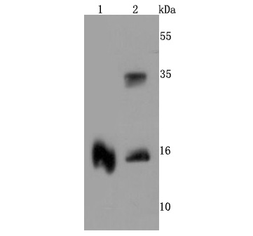
Fig1: Western blot analysis of TTR on different cells lysates using anti-TTR antibody at 1/500 dilution. Positive control: Line 1: Human brain Line 2;Human lungApplication
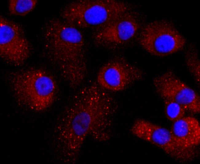
Fig2: ICC staining TTR in A549 cells (red). The nuclear counter stain is DAPI (blue). Cells were fixed in paraformaldehyde, permeabilised with 0.25% Triton X100/PBS.Application
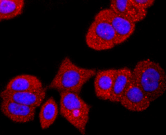
Fig3: ICC staining TTR in MCF-7 cells (red). The nuclear counter stain is DAPI (blue). Cells were fixed in paraformaldehyde, permeabilised with 0.25% Triton X100/PBS.Application
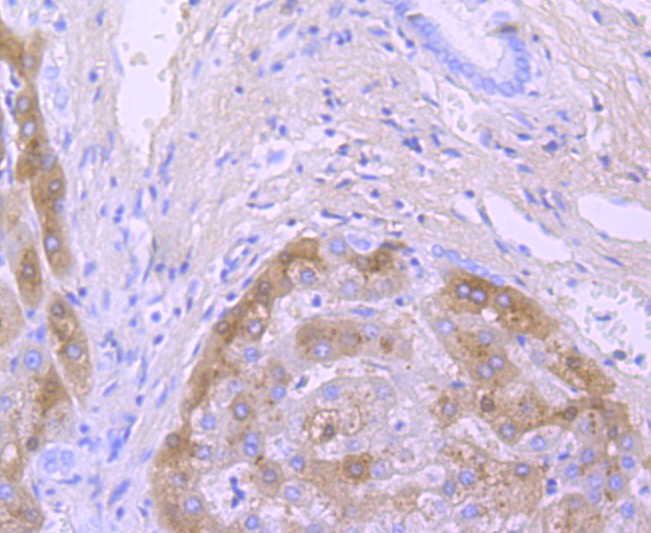
Fig4: Immunohistochemical analysis of paraffin-embedded human liver cancer tissue using anti-TTR antibody. Counter stained with hematoxylin.Application
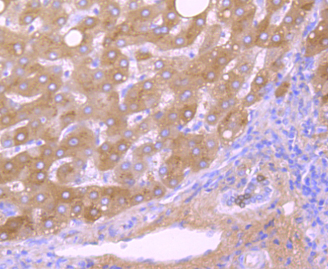
Fig5: Immunohistochemical analysis of paraffin-embedded human liver tissue using anti- TTR antibody. Counter stained with hematoxylin.Application
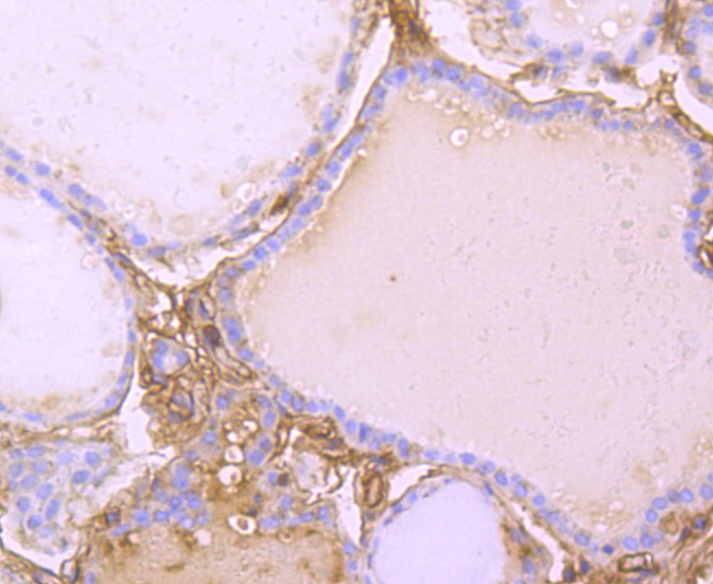
Fig6: Immunohistochemical analysis of paraffin-embedded human thyroid tissue using anti-TTR antibody. Counter stained with hematoxylin.Application
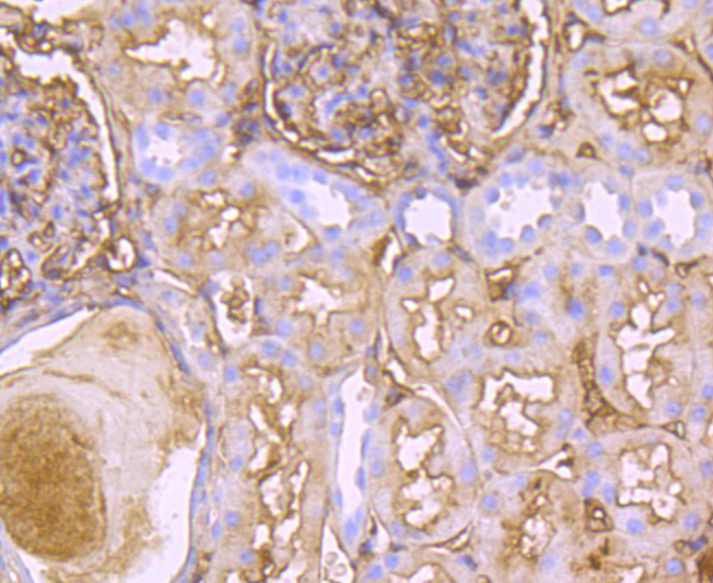
Fig7: Immunohistochemical analysis of paraffin-embedded human kidney tissue using anti-TTR antibody. Counter stained with hematoxylin.Application
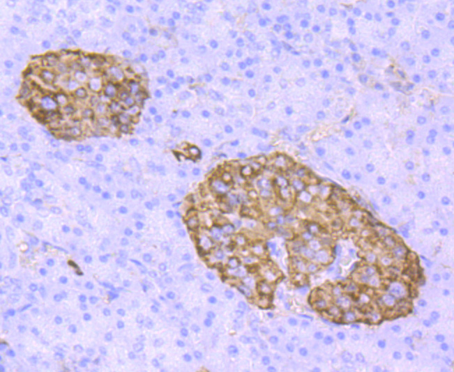
Fig8: Immunohistochemical analysis of paraffin-embedded human pancreas tissue using anti-TTR antibody. Counter stained with hematoxylin.Application
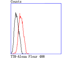
Fig9: Flow cytometric analysis of MCF-7 cells with TTR antibody at 1/50 dilution (red) compared with an unlabelled control (cells without incubation with primary antibody; black). Alexa Fluor 488-conjugated goat anti rabbit IgG was used as the secondary antibody.| Product Name | TTR [JM11-43] |
|---|---|
| Antibody Type | Primary Antibodies |
| Antigen Alias | Alternative names Amyloid polyneuropathy antibody Amyloidosis I antibody ATTR antibody Carpal tunnel syndrome 1 antibody CTS antibody CTS1 antibody Dysprealbuminemic euthyroidal hyperthyroxinemia antibody Dystransthyretinemic hyperthyroxinemia antibody Epididymis luminal protein 111 antibody HEL111 antibody HsT2651 antibody PALB antibody Prealbumin amyloidosis type I antibody Prealbumin antibody Prealbumin Thyroxine-binding antibody Senile systemic amyloidosis antibody TBPA antibody Thyroxine binding prealbumin antibody Transthyretin antibody TTHY_HUMAN antibody TTR antibody TTR protein antibod |
| Product description | Prealbumin, also designated transthyretin, is a major thyroid-hormone binding protein involved in transporting thyroxine from the bloodstream to the brain. Prealbumin is located in the cytoplasm and in the vesicles of developing rat brain cells, and is thought to be transported there from the cerebrospinal fluid via endocytosis. Sequence variants of this protein have been identified in amyloid fibrils from patients with familial amyloidotic polyneuropathy (FAP), the most common form of hereditary systemic amyloidosis. Although the biologically active form of Prealbumin is a tetramer, the amyloidogenic intermed-iate is thought to be a monomeric species. Prealbumin also binds to the retinol carrier protein RBP (retinol-binding protein). The gene encoding Prealbumin maps to human chromosome 18q12.1. |
| Immunogen | recombinant protein |
| Clonality | Monoclonal |
|---|---|
| Isotype | IgG |
| Host Species | Recombinant rabbit |
| Tested Applications | WBIPICC/IFIHCFC |
| WB:1:500-1:2,000 ICC:1:50-1:200 IHC:1:50-1:200 FC:1:50-1:100 |
|
| Species Reactivity | Human |
| Concentration | 1mg/ml |
| Alternative Names | Alternative names Amyloid polyneuropathy antibody Amyloidosis I antibody ATTR antibody Carpal tunnel syndrome 1 antibody CTS antibody CTS1 antibody Dysprealbuminemic euthyroidal hyperthyroxinemia antibody Dystransthyretinemic hyperthyroxinemia antibody Epididymis luminal protein 111 antibody HEL111 antibody HsT2651 antibody PALB antibody Prealbumin amyloidosis type I antibody Prealbumin antibody Prealbumin Thyroxine-binding antibody Senile systemic amyloidosis antibody TBPA antibody Thyroxine binding prealbumin antibody Transthyretin antibody TTHY_HUMAN antibody TTR antibody TTR protein antibod |
|---|---|
| Molecular Weight(MW) | 16 kDa |
| Cellular Localization | Secreted.Cytoplasm. |
| SwissProt ID | P02766 |
|---|
Application
Fig1: Western blot analysis of TTR on different cells lysates using anti-TTR antibody at 1/500 dilution. Positive control: Line 1: Human brain Line 2;Human lung
Application
Fig2: ICC staining TTR in A549 cells (red). The nuclear counter stain is DAPI (blue). Cells were fixed in paraformaldehyde, permeabilised with 0.25% Triton X100/PBS.
Application
Fig3: ICC staining TTR in MCF-7 cells (red). The nuclear counter stain is DAPI (blue). Cells were fixed in paraformaldehyde, permeabilised with 0.25% Triton X100/PBS.
Application
Fig4: Immunohistochemical analysis of paraffin-embedded human liver cancer tissue using anti-TTR antibody. Counter stained with hematoxylin.
Application
Fig5: Immunohistochemical analysis of paraffin-embedded human liver tissue using anti- TTR antibody. Counter stained with hematoxylin.
Application
Fig6: Immunohistochemical analysis of paraffin-embedded human thyroid tissue using anti-TTR antibody. Counter stained with hematoxylin.
Application
Fig7: Immunohistochemical analysis of paraffin-embedded human kidney tissue using anti-TTR antibody. Counter stained with hematoxylin.
Application
Fig8: Immunohistochemical analysis of paraffin-embedded human pancreas tissue using anti-TTR antibody. Counter stained with hematoxylin.
Application
Fig9: Flow cytometric analysis of MCF-7 cells with TTR antibody at 1/50 dilution (red) compared with an unlabelled control (cells without incubation with primary antibody; black). Alexa Fluor 488-conjugated goat anti rabbit IgG was used as the secondary antibody.| Positive Control | Human brain, human lung, MCF-7, A549, Hela, human liver tissue, human kidney tissue, human liver cancer tissue, human thyroid tissue, human pancreas tissue. |
|---|---|
| Application Notes | WB:1:500-1:2,000 ICC:1:50-1:200 IHC:1:50-1:200 FC:1:50-1:100 |
| Form | Liquid |
|---|---|
| Storage Instructions | Store at +4℃ after thawing. Aliquot store at -20℃ or -80℃. Avoid repeated freeze / thaw cycles. |
| Storage Buffer | 1*TBS (pH7.4), 1%BSA, 40%Glycerol. Preservative: 0.05% Sodium Azide. |


