Application
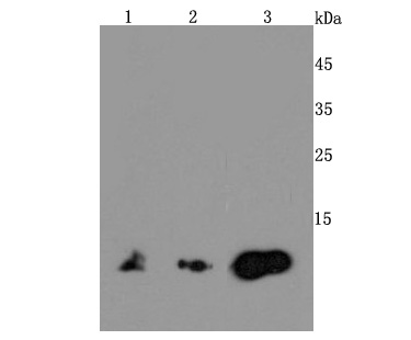
Fig1: Western blot analysis of MIF on different cells lysates using anti-MIF antibody at 1/500 dilution. Positive control: Line 1: Mouse brain Line 2: Jurkat Line 3: HelaApplication
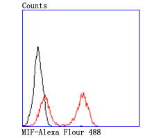
Fig2: Flow cytometric analysis of THP-1 cells with MIF antibody at 1/50 dilution (red) compared with an unlabelled control (cells without incubation with primary antibody; black). Alexa Fluor 488-conjugated goat anti rabbit IgG was used as the secondary antibody.| Product Name | MIF [JM11-64] |
|---|---|
| Antibody Type | Primary Antibodies |
| Antigen Alias | GIF antibody GLIF antibody Glycosylation inhibiting factor antibody Glycosylation-inhibiting factor antibody L-dopachrome isomerase antibody L-dopachrome tautomerase antibody Macrophage migration inhibitory factor (glycosylation-inhibiting factor) antibody Macrophage migration inhibitory factor antibody MIF antibody MIF protein antibody MIF_HUMAN antibody MMIF antibody Phenylpyruvate tautomerase antibody |
| Product description | ponse to infection. MIF is expressed at sites of inflammation, which suggests that it plays a role in regulating macrophage function in host defense. MIF is produced by the pituitary gland and is found in monocytes, macrophages, differentiating immunological cells in the eye lens and brain, and fibroblasts. Elevated levels of MIF protein are detected in the plasma of patients with severe sepsis or septic shock, a condition where MIF influences endotoxic shock by enhancing the production of other inflammatory cytokines including tumor necrosis factor α (TNFα), interleukin-1 (IL-1) and interferon-γ (IFN-γ). MIF promotes the systemic inflammatory response by counter-regulating glucocorticoid-mediated inhibition of immune-cell activation and proinflammatory cytokine production. MIF may mediate tissue destruction through the induction of proteinases. |
| Immunogen | recombinant protein |
| Clonality | Monoclonal |
|---|---|
| Isotype | IgG |
| Host Species | Recombinant rabbit |
| Tested Applications | WBIPFC |
| WB:1:500-1:2,000 IP:1:10-1:50 FC:1:50-1:100 |
|
| Species Reactivity | HumanMouseRat |
| Concentration | 1mg/ml |
| Alternative Names | GIF antibody GLIF antibody Glycosylation inhibiting factor antibody Glycosylation-inhibiting factor antibody L-dopachrome isomerase antibody L-dopachrome tautomerase antibody Macrophage migration inhibitory factor (glycosylation-inhibiting factor) antibody Macrophage migration inhibitory factor antibody MIF antibody MIF protein antibody MIF_HUMAN antibody MMIF antibody Phenylpyruvate tautomerase antibody |
|---|---|
| Molecular Weight(MW) | 12 kDa |
| Cellular Localization | Secreted. Cytoplasm. |
Application
Fig1: Western blot analysis of MIF on different cells lysates using anti-MIF antibody at 1/500 dilution. Positive control: Line 1: Mouse brain Line 2: Jurkat Line 3: Hela
Application
Fig2: Flow cytometric analysis of THP-1 cells with MIF antibody at 1/50 dilution (red) compared with an unlabelled control (cells without incubation with primary antibody; black). Alexa Fluor 488-conjugated goat anti rabbit IgG was used as the secondary antibody.| Positive Control | Jurkat, Hela, THP-1, mouse brain. |
|---|---|
| Application Notes | WB:1:500-1:2,000 IP:1:10-1:50 FC:1:50-1:100 |
| Form | Liquid |
|---|---|
| Storage Instructions | Store at +4℃ after thawing. Aliquot store at -20℃ or -80℃. Avoid repeated freeze / thaw cycles. |
| Storage Buffer | 1*TBS (pH7.4), 1%BSA, 40%Glycerol. Preservative: 0.05% Sodium Azide. |


