Application
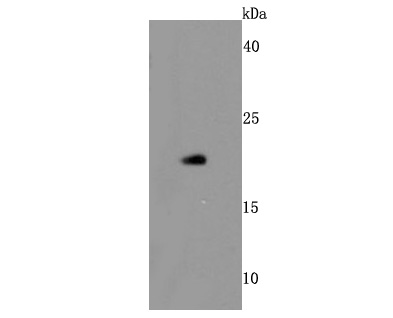
Fig1: Western blot analysis of Bid on human thymus cells lysates using anti-Bid antibody at 1/500 dilution.Application
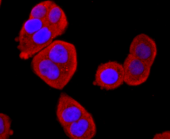
Fig2: ICC staining Bid in Hela cells (red). The nuclear counter stain is DAPI (blue). Cells were fixed in paraformaldehyde, permeabilised with 0.25% Triton X100/PBS.Application
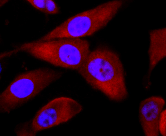
Fig3: ICC staining Bid in PC-3M cells (red). The nuclear counter stain is DAPI (blue). Cells were fixed in paraformaldehyde, permeabilised with 0.25% Triton X100/PBS.Application
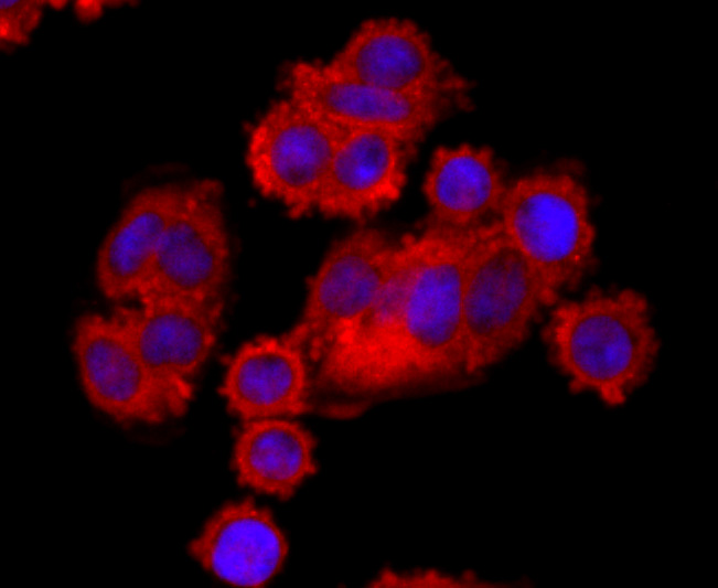
Fig4: ICC staining Bid in SW480 cells (red). The nuclear counter stain is DAPI (blue). Cells were fixed in paraformaldehyde, permeabilised with 0.25% Triton X100/PBS.Application
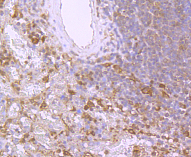
Fig5: Immunohistochemical analysis of paraffin-embedded human tonsil tissue using anti-Bid antibody. Counter stained with hematoxylin.Application
Fig6: Immunohistochemical analysis of paraffin-embedded human lung tissue using anti-Bid antibody. Counter stained with hematoxylin.Application
Fig7: Immunohistochemical analysis of paraffin-embedded human liver cancer tissue using anti-Bid antibody. Counter stained with hematoxylin.Application
Fig8: Immunohistochemical analysis of paraffin-embedded human colon cancer tissue using anti-Bid antibody. Counter stained with hematoxylin.Application
Fig9: Immunohistochemical analysis of paraffin-embedded human spleen tissue using anti-Bid antibody. Counter stained with hematoxylin.Application
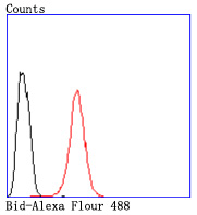
Fig10: Flow cytometric analysis of Hela cells with Bid antibody at 1/50 dilution (red) compared with an unlabelled control (cells without incubation with primary antibody; black). Alexa Fluor 488-conjugated goat anti rabbit IgG was used as the secondary| Product Name | Bid [JM11-14] |
|---|---|
| Antibody Type | Primary Antibodies |
| Antigen Alias | Apoptic death agonist antibody Apoptotic death agonist BID antibody BH3 interacting domain death agonist antibody BH3 interacting domain death agonist p11 antibody BH3 interacting domain death agonist p13 antibody BH3 interacting domain death agonist p15 antibody BH3-interacting domain death agonist p11 antibody BID antibody BID isoform ES(1b) antibody BID isoform L(2) antibody BID isoform Si6 antibody BID_HUMAN antibody Desmocollin type 4 antibody FP497 antibody Human BID coding sequence antibody MGC15319 antibody MGC42355 antibody p11 BID antibody p13 BID antibody p15 BID antibody p22 BID antibody |
| Product description | Members of the Bcl-2 family of proteins interact to regulate programmed cell death, or apoptosis. Various homodimers and heterodimers formed by proteins in this family can either promote or inhibit apoptosis. Bcl-2 blocks cell death following a variety of stimuli and confers a death-sparing effect on certain hematopoietic cell lines following growth factor withdrawal. Additional apoptotic inhibitors in this family include A1, Bag-1, Bcl-w, Bcl-x and Mcl-1. Pro-apoptotic members of this family include Bax, Bad, Bak, Bik (NBK) and BID. BID contains a BH3 domain which allows it to dimerize with and counter the death repressor effects of Bcl-2. BID has also been shown to heterodimerize with Bcl-x and the death agonist Bax. BID is localized predominantly in the cytosol and is also present in membrane fractions. It is highly expressed in kidney and can also be detected in brain, spleen, liver, testis and lung. |
| Immunogen | recombinant protein |
| Clonality | Monoclonal |
|---|---|
| Isotype | IgG |
| Host Species | Recombinant rabbit |
| Tested Applications | WBICC/IFIHCFCIP |
| WB:1:500-1:1,000 ICC:1:50-1:200 IHC:1:50-1:200 FC:1:50-1:100 IP:1:10-1:50 |
|
| Species Reactivity | Human |
| Concentration | 1mg/ml |
| Alternative Names | Apoptic death agonist antibody Apoptotic death agonist BID antibody BH3 interacting domain death agonist antibody BH3 interacting domain death agonist p11 antibody BH3 interacting domain death agonist p13 antibody BH3 interacting domain death agonist p15 antibody BH3-interacting domain death agonist p11 antibody BID antibody BID isoform ES(1b) antibody BID isoform L(2) antibody BID isoform Si6 antibody BID_HUMAN antibody Desmocollin type 4 antibody FP497 antibody Human BID coding sequence antibody MGC15319 antibody MGC42355 antibody p11 BID antibody p13 BID antibody p15 BID antibody p22 BID antibody |
|---|---|
| Molecular Weight(MW) | 22 kDa |
| Cellular Localization | Cytoplasm. Mitochondrion membrane. |
| SwissProt ID | P55957 |
|---|
Application
Fig1: Western blot analysis of Bid on human thymus cells lysates using anti-Bid antibody at 1/500 dilution.
Application
Fig2: ICC staining Bid in Hela cells (red). The nuclear counter stain is DAPI (blue). Cells were fixed in paraformaldehyde, permeabilised with 0.25% Triton X100/PBS.
Application
Fig3: ICC staining Bid in PC-3M cells (red). The nuclear counter stain is DAPI (blue). Cells were fixed in paraformaldehyde, permeabilised with 0.25% Triton X100/PBS.
Application
Fig4: ICC staining Bid in SW480 cells (red). The nuclear counter stain is DAPI (blue). Cells were fixed in paraformaldehyde, permeabilised with 0.25% Triton X100/PBS.
Application
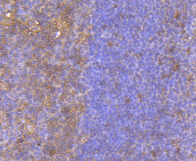
Fig5: Immunohistochemical analysis of paraffin-embedded human tonsil tissue using anti-Bid antibody. Counter stained with hematoxylin.
Application
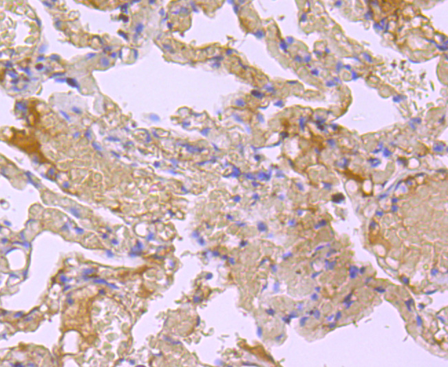
Fig6: Immunohistochemical analysis of paraffin-embedded human lung tissue using anti-Bid antibody. Counter stained with hematoxylin.
Application
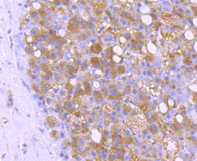
Fig7: Immunohistochemical analysis of paraffin-embedded human liver cancer tissue using anti-Bid antibody. Counter stained with hematoxylin.
Application
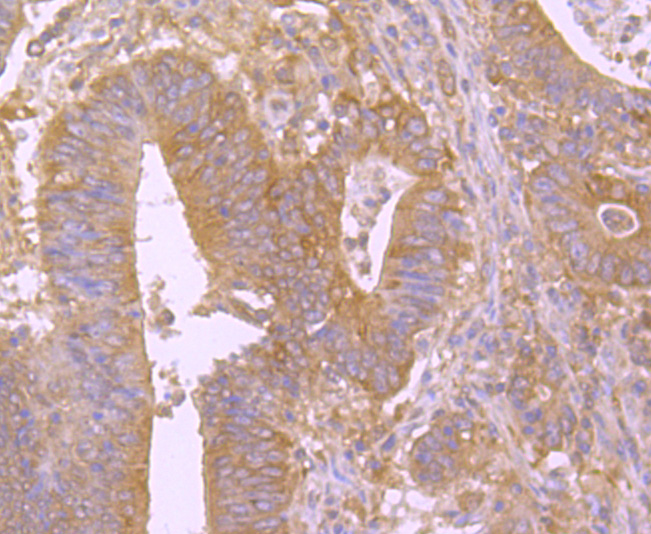
Fig8: Immunohistochemical analysis of paraffin-embedded human colon cancer tissue using anti-Bid antibody. Counter stained with hematoxylin.
Application
Fig9: Immunohistochemical analysis of paraffin-embedded human spleen tissue using anti-Bid antibody. Counter stained with hematoxylin.
Application
Fig10: Flow cytometric analysis of Hela cells with Bid antibody at 1/50 dilution (red) compared with an unlabelled control (cells without incubation with primary antibody; black). Alexa Fluor 488-conjugated goat anti rabbit IgG was used as the secondary| Positive Control | HepG2, SW480, Hela, human thymus, human tonsil tissue, human lung tissue, human liver cancer tissue, human colon cancer tissue, human spleen tissue. |
|---|---|
| Application Notes | WB:1:500-1:1,000 ICC:1:50-1:200 IHC:1:50-1:200 FC:1:50-1:100 IP:1:10-1:50 |
| Form | Liquid |
|---|---|
| Storage Instructions | Store at +4℃ after thawing. Aliquot store at -20℃ or -80℃. Avoid repeated freeze / thaw cycles. |
| Storage Buffer | 1*TBS (pH7.4), 1%BSA, 40%Glycerol. Preservative: 0.05% Sodium Azide. |


