Application
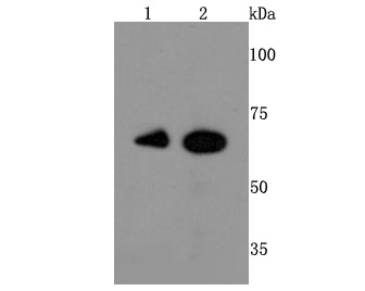
Fig1: Western blot analysis of Ku 70 on different cells lysates using anti-Ku 70 antibody at 1/500 dilution. Positive control: Line 1: Hela Line 2: JurkatApplication
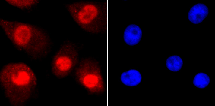
Fig2: ICC staining Ku 70 in A549 cells (red). The nuclear counter stain is DAPI (blue). Cells were fixed in paraformaldehyde, permeabilised with 0.25% Triton X100/PBS.Application
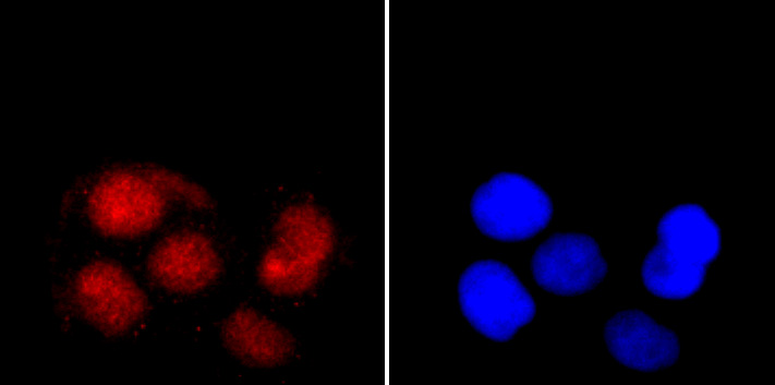
Fig3: ICC staining Ku 70 in Hela cells (red). The nuclear counter stain is DAPI (blue). Cells were fixed in paraformaldehyde, permeabilised with 0.25% Triton X100/PBS.Application
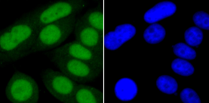
Fig4: ICC staining Ku 70 in MCF-7 cells (green). The nuclear counter stain is DAPI (blue). Cells were fixed in paraformaldehyde, permeabilised with 0.25% Triton X100/PBS.Application
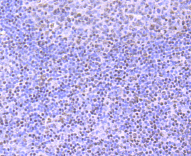
Fig5: Immunohistochemical analysis of paraffin-embedded human tonsil tissue using anti-Ku 70 antibody. Counter stained with hematoxylin.Application
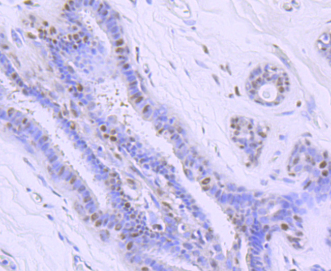
Fig6: Immunohistochemical analysis of paraffin-embedded human breast cancer tissue using anti-Ku 70 antibody. Counter stained with hematoxylin.Application
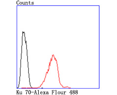
Fig7: Flow cytometric analysis of Hela cells with Ku 70 antibody at 1/50 dilution (red) compared with an unlabelled control (cells without incubation with primary antibody; black). Alexa Fluor 488-conjugated goat anti rabbit IgG was used as the secondary antibody.| Product Name | Ku 70 [JM61-31] |
|---|---|
| Antibody Type | Primary Antibodies |
| Antigen Alias | 5''-deoxyribose-5-phosphate lyase Ku70 antibody 5''-dRP lyase Ku70 antibody 70 kDa subunit of Ku antigen antibody ATP dependent DNA helicase 2 subunit 1 antibody ATP dependent DNA helicase II 70 kDa subunit antibody ATP-dependent DNA helicase 2 subunit 1 antibody ATP-dependent DNA helicase II 70 kDa subunit antibody CTC box binding factor 75 kDa subunit antibody CTC box-binding factor 75 kDa subunit antibody CTC75 antibody CTCBF antibody DNA repair protein XRCC6 antibody G22P1 antibody Ku 70 antibody Ku autoantigen p70 subunit antibody Ku autoantigen, 70kDa antibody Ku p70 antibody Ku70 antibody Ku70 DNA binding component of DNA-dependent proteinkinase complex (thyroid autoantigen 70 kDa antibody Kup70 antibody Lupus Ku autoantigen protein p70 antibody ML8 antibody Thyroid autoantigen 70kD (Ku antigen) antibody Thyroid autoantigen antibody Thyroid lupus autoantigen antibody Thyroid lupus autoantigen p70 antibody Thyroid-lupus autoantigen antibody TLAA antibody X ray repair complementing defective repair in Chinese hamster cells 6 antibody X-ray repair complementing defective repair in Chinese hamster cells 6 antibody X-ray repair cross-complementing protein 6 antibody XRCC 6 antibody Xrcc6 antibody XRCC6_HUMAN antibody |
| Product description | The Ku protein is localized in the nucleus and is composed of subunits referred to as Ku-70 (p70) and Ku-86 (p86) which is also known by the synonym Ku-80 or (p80). Ku was first described as an autoantigen to which antibodies were produced in a patient with scleroderma polymyositis overlap syndrome, and was later found in the sera of patients with other rheumatic diseases. Both subunits of the Ku protein have been cloned, and a number of functions have been proposed for Ku, including cell signaling, DNA replication and transcriptional activation. Ku is involved in Pol II-directed transcription by virtue of its DNA binding activity, serving as the regulatory component of the DNA-associated protein kinase that phosphorylates Pol II and transcription factor Sp. Ku proteins also activate transcription from the U1 small nuclear RNA and the human transferrin receptor gene promoters. A Ku-related protein designated the enhancer 1 binding factor (E1BF), composed of two subunits, has been identified as a positive regulator of RNA polymerase I transcription initiation. |
| Immunogen | recombinant protein |
| Clonality | Monoclonal |
|---|---|
| Isotype | IgG |
| Host Species | Recombinant rabbit |
| Tested Applications | WBIPICC/IFIHCFC |
| WB:1:500-1:2,000 ICC:1:50-1:200 IHC:1:50-1:200 FC:1:50-1:100 IP:1:10-1:50 |
|
| Species Reactivity | Human |
| Concentration | 1mg/ml |
| Alternative Names | 5''-deoxyribose-5-phosphate lyase Ku70 antibody
5''-dRP lyase Ku70 antibody
70 kDa subunit of Ku antigen antibody
ATP dependent DNA helicase 2 subunit 1 antibody
ATP dependent DNA helicase II 70 kDa subunit antibody
ATP-dependent DNA helicase 2 subunit 1 antibody
ATP-dependent DNA helicase II 70 kDa subunit antibody
CTC box binding factor 75 kDa subunit antibody
CTC box-binding factor 75 kDa subunit antibody
CTC75 antibody
CTCBF antibody
DNA repair protein XRCC6 antibody
G22P1 antibody
Ku 70 antibody
Ku autoantigen p70 subunit antibody
Ku autoantigen 70kDa antibody Ku p70 antibody Ku70 antibody Ku70 DNA binding component of DNA-dependent proteinkinase complex (thyroid autoantigen 70 kDa antibody Kup70 antibody Lupus Ku autoantigen protein p70 antibody ML8 antibody Thyroid autoantigen 70kD (Ku antigen) antibody Thyroid autoantigen antibody Thyroid lupus autoantigen antibody Thyroid lupus autoantigen p70 antibody Thyroid-lupus autoantigen antibody TLAA antibody X ray repair complementing defective repair in Chinese hamster cells 6 antibody X-ray repair complementing defective repair in Chinese hamster cells 6 antibody X-ray repair cross-complementing protein 6 antibody XRCC 6 antibody Xrcc6 antibody XRCC6_HUMAN antibody |
|---|---|
| Molecular Weight(MW) | 70 kDa |
| Cellular Localization | Nucleus. Chromosome. |
| SwissProt ID | P12956 |
|---|
Application
Fig1: Western blot analysis of Ku 70 on different cells lysates using anti-Ku 70 antibody at 1/500 dilution. Positive control: Line 1: Hela Line 2: Jurkat
Application
Fig2: ICC staining Ku 70 in A549 cells (red). The nuclear counter stain is DAPI (blue). Cells were fixed in paraformaldehyde, permeabilised with 0.25% Triton X100/PBS.
Application
Fig3: ICC staining Ku 70 in Hela cells (red). The nuclear counter stain is DAPI (blue). Cells were fixed in paraformaldehyde, permeabilised with 0.25% Triton X100/PBS.
Application
Fig4: ICC staining Ku 70 in MCF-7 cells (green). The nuclear counter stain is DAPI (blue). Cells were fixed in paraformaldehyde, permeabilised with 0.25% Triton X100/PBS.
Application
Fig5: Immunohistochemical analysis of paraffin-embedded human tonsil tissue using anti-Ku 70 antibody. Counter stained with hematoxylin.
Application
Fig6: Immunohistochemical analysis of paraffin-embedded human breast cancer tissue using anti-Ku 70 antibody. Counter stained with hematoxylin.
Application
Fig7: Flow cytometric analysis of Hela cells with Ku 70 antibody at 1/50 dilution (red) compared with an unlabelled control (cells without incubation with primary antibody; black). Alexa Fluor 488-conjugated goat anti rabbit IgG was used as the secondary antibody.| Positive Control | Jurkat, A549, MCF-7, Hela, human tonsil tissue, human breast cancer tissue. |
|---|---|
| Application Notes | WB:1:500-1:2,000 ICC:1:50-1:200 IHC:1:50-1:200 FC:1:50-1:100 IP:1:10-1:50 |
| Form | Liquid |
|---|---|
| Storage Instructions | Store at +4℃ after thawing. Aliquot store at -20℃ or -80℃. Avoid repeated freeze / thaw cycles. |
| Storage Buffer | 1*TBS (pH7.4), 1%BSA, 40%Glycerol. Preservative: 0.05% Sodium Azide. |


