Application
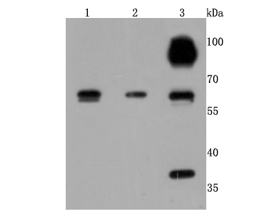
Fig1: Western blot analysis of Cathepsin B on different cells lysates using anti-Cathepsin B antibody at 1/500 dilution. Positive control: Lane 1: HepG2 Lane 2: Hela Lane 3:A549Application
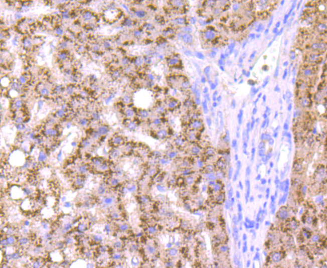
Fig2: Immunohistochemical analysis of paraffin-embedded human liver cancer tissue using anti-Cathepsin B antibody. Counter stained with hematoxylin.Application
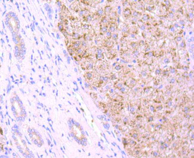
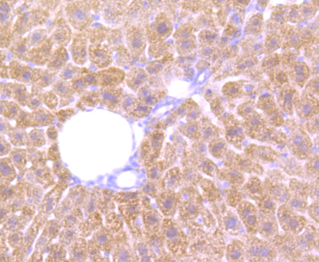
Fig3: Immunohistochemical analysis of paraffin-embedded human liver tissue using anti-Cathepsin B antibody. Counter stained with hematoxylin.Application
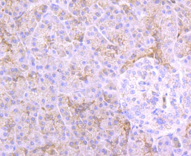
Fig4: Immunohistochemical analysis of paraffin-embedded human pancreas tissue using anti-Cathepsin B antibody. Counter stained with hematoxylin.Application
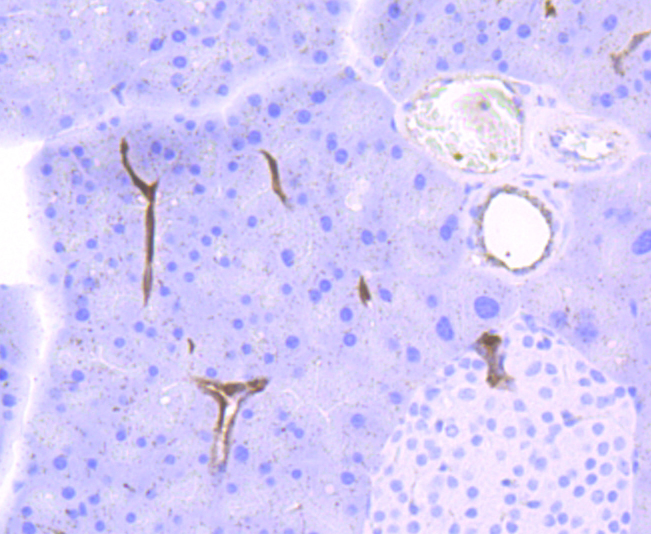
Fig5: Immunohistochemical analysis of paraffin-embedded mouse liver tissue using anti-Cathepsin B antibody. Counter stained with hematoxylin.Application
Fig6: Immunohistochemical analysis of paraffin-embedded mouse pancreas tissue using anti-Cathepsin B antibody. Counter stained with hematoxylin.Application
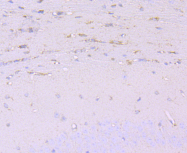
Fig7: Immunohistochemical analysis of paraffin-embedded mouse brain tissue using anti-Cathepsin B antibody. Counter stained with hematoxylin.| Product Name | Cathepsin B [JA11-02] |
|---|---|
| Antibody Type | Primary Antibodies |
| Antigen Alias | Amyloid precursor protein secretase antibody APP secretase antibody APPS antibody CATB_HUMAN antibody Cathepsin B heavy chain antibody Cathepsin B1 antibody CathepsinB antibody CPSB antibody CTSB antibody cysteine protease antibody OTTHUMP00000116009 antibody OTTHUMP00000229510 antibody OTTHUMP00000229511 antibody OTTHUMP00000229512 antibody OTTHUMP00000229514 antibody OTTHUMP00000229515 antibody OTTHUMP00000229516 antibody Preprocathepsin B antibody |
| Product description | The cathepsin family of proteolytic enzymes contains several diverse classes of proteases. The cysteine protease class comprises cathepsins B, L, H, K, S, and O. The aspartyl protease class is composed of cathepsins D and E. Cathepsin G is in the serine protease class. Most cathepsins are lysosomal and each is involved in cellular metabolism, participating in various events such as peptide biosynthesis and protein degradation. Cathepsin B is expressed in luminal epithelial cells, indicating that cathepsin B is a marker for secretory cell death. |
| Immunogen | recombinant protein |
| Clonality | Monoclonal |
|---|---|
| Isotype | IgG |
| Host Species | Recombinant rabbit |
| Tested Applications | WBICC/IFIHC |
| WB:1:500-1:1,000 ICC:1:50-1:200 IHC:1:50-1:200 |
|
| Species Reactivity | Human |
| Concentration | 1mg/ml |
| Alternative Names | Amyloid precursor protein secretase antibody APP secretase antibody APPS antibody CATB_HUMAN antibody Cathepsin B heavy chain antibody Cathepsin B1 antibody CathepsinB antibody CPSB antibody CTSB antibody cysteine protease antibody OTTHUMP00000116009 antibody OTTHUMP00000229510 antibody OTTHUMP00000229511 antibody OTTHUMP00000229512 antibody OTTHUMP00000229514 antibody OTTHUMP00000229515 antibody OTTHUMP00000229516 antibody Preprocathepsin B antibody |
|---|---|
| Molecular Weight(MW) | 38 kDa |
| Cellular Localization | Lysosome. Melanosome. |
| SwissProt ID | P07858 |
|---|
Application
Fig1: Western blot analysis of Cathepsin B on different cells lysates using anti-Cathepsin B antibody at 1/500 dilution. Positive control: Lane 1: HepG2 Lane 2: Hela Lane 3:A549
Application
Fig2: Immunohistochemical analysis of paraffin-embedded human liver cancer tissue using anti-Cathepsin B antibody. Counter stained with hematoxylin.
Application
Fig3: Immunohistochemical analysis of paraffin-embedded human liver tissue using anti-Cathepsin B antibody. Counter stained with hematoxylin.
Application
Fig4: Immunohistochemical analysis of paraffin-embedded human pancreas tissue using anti-Cathepsin B antibody. Counter stained with hematoxylin.
Application
Fig5: Immunohistochemical analysis of paraffin-embedded mouse liver tissue using anti-Cathepsin B antibody. Counter stained with hematoxylin.
Application
Fig6: Immunohistochemical analysis of paraffin-embedded mouse pancreas tissue using anti-Cathepsin B antibody. Counter stained with hematoxylin.
Application
Fig7: Immunohistochemical analysis of paraffin-embedded mouse brain tissue using anti-Cathepsin B antibody. Counter stained with hematoxylin.| Positive Control | HepG2, A549, Hela, human liver tissue, human liver cancer tissue, human pancreas tissue, mouse pancreas tissue, mouse brain tissue. |
|---|---|
| Application Notes | WB:1:500-1:1,000 ICC:1:50-1:200 IHC:1:50-1:200 |
| Form | Liquid |
|---|---|
| Storage Instructions | Store at +4℃ after thawing. Aliquot store at -20℃ or -80℃. Avoid repeated freeze / thaw cycles. |
| Storage Buffer | 1*TBS (pH7.4), 1%BSA, 40%Glycerol. Preservative: 0.05% Sodium Azide. |


