WB
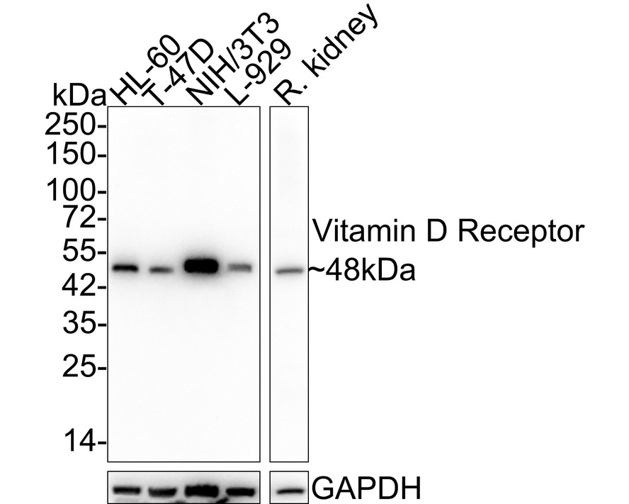
Western blot analysis of Vitamin D Receptor on different lysates with Rabbit anti-Vitamin D Receptor antibody at 1/5,000 dilution. Lane 1: HL-60 cell lysate (15 µg/Lane), Lane 2: T-47D cell lysate (15 µg/Lane), Lane 3: NIH/3T3 cell lysate (15 µg/Lane), Lane 4: L-929 cell lysate (15 µg/Lane), Lane 5: Rat kidney tissue lysate (20 µg/Lane), Exposure time: 30 seconds; 4-20% SDS-PAGE gel. Proteins were transferred to a PVDF membrane and blocked with 5% NFDM/TBST for 1 hour at room temperature. The primary antibody at 1/5,000 dilution was used in 5% NFDM/TBST at 4℃ overnight. Goat Anti-Rabbit IgG - HRP Secondary Antibody at 1/50,000 dilution was used for 1 hour at room temperature.IHC
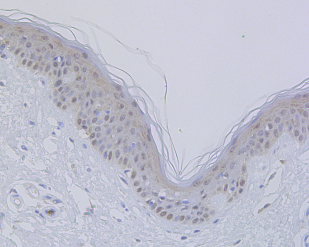
Immunohistochemical analysis of paraffin-embedded human skin tissue using anti-Vitamin D Receptor antibody. The section was pre-treated using heat mediated antigen retrieval with sodium citrate buffer (pH 6.0) for 20 minutes. The tissues were blocked in 5% BSA for 30 minutes at room temperature, washed with ddH2O and PBS, and then probed with the antibody (ET1704-09) at 1/100 dilution, for 30 minutes at room temperature and detected using an HRP conjugated compact polymer system. DAB was used as the chromogen. Counter stained with hematoxylin and mounted with DPX.IHC
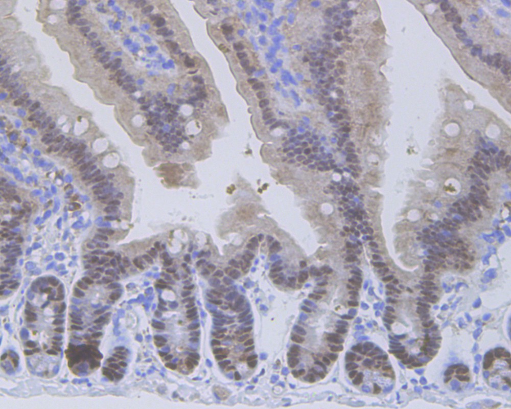
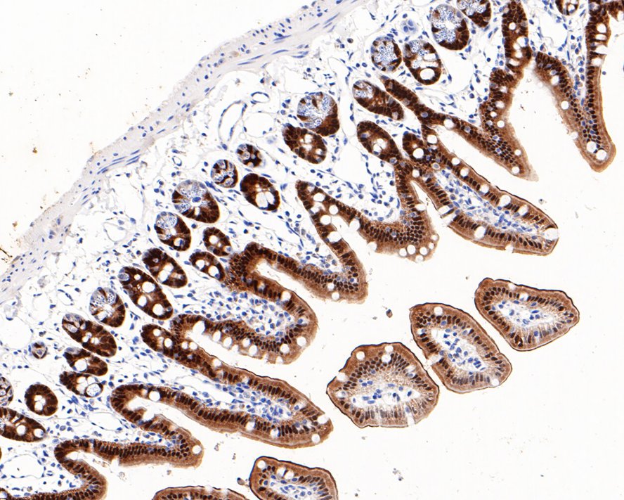
Immunohistochemical analysis of paraffin-embedded mouse colon tissue using anti-Vitamin D Receptor antibody. The section was pre-treated using heat mediated antigen retrieval with sodium citrate buffer (pH 6.0) for 20 minutes. The tissues were blocked in 5% BSA for 30 minutes at room temperature, washed with ddH2O and PBS, and then probed with the antibody (ET1704-09) at 1/100 dilution, for 30 minutes at room temperature and detected using an HRP conjugated compact polymer system. DAB was used as the chromogen. Counter stained with hematoxylin and mounted with DPX.IHC
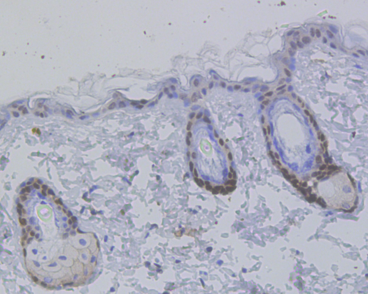
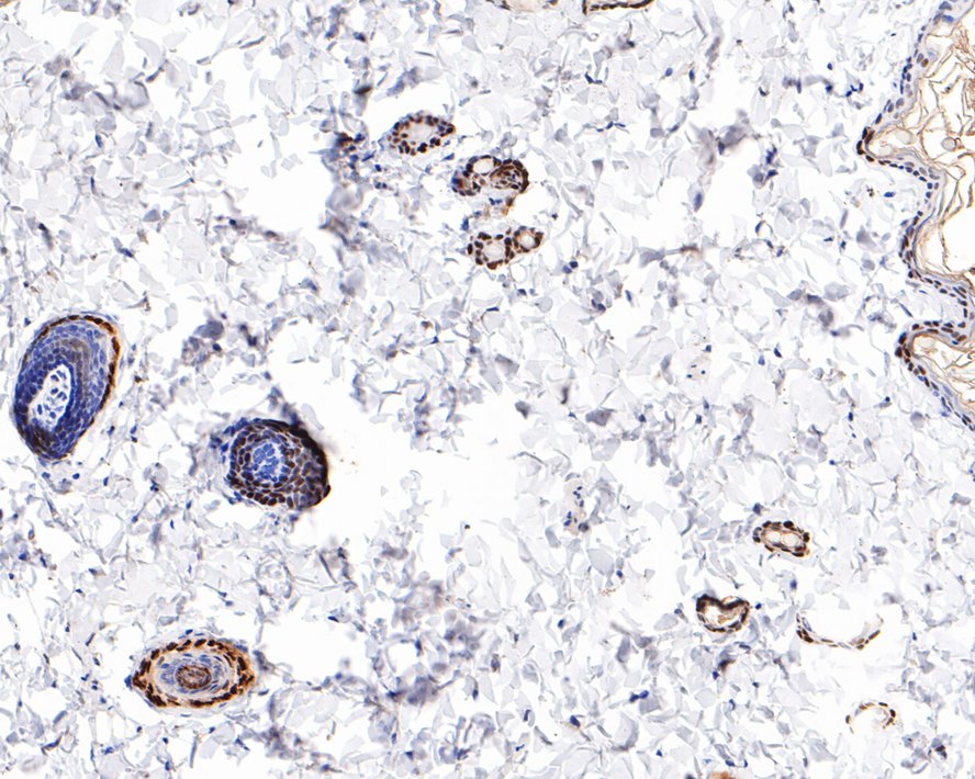
Immunohistochemical analysis of paraffin-embedded mouse skin tissue using anti-Vitamin D Receptor antibody. The section was pre-treated using heat mediated antigen retrieval with sodium citrate buffer (pH 6.0) for 20 minutes. The tissues were blocked in 5% BSA for 30 minutes at room temperature, washed with ddH2O and PBS, and then probed with the antibody (ET1704-09) at 1/100 dilution, for 30 minutes at room temperature and detected using an HRP conjugated compact polymer system. DAB was used as the chromogen. Counter stained with hematoxylin and mounted with DPX.IHC
Immunohistochemical analysis of paraffin-embedded mouse colon tissue with Rabbit anti-Vitamin D Receptor antibody at 1/800 dilution. The section was pre-treated using heat mediated antigen retrieval with sodium citrate buffer (pH 6.0) (high pressure) for 2 minutes. The tissues were blocked in 1% BSA for 20 minutes at room temperature, washed with ddH2O and PBS, and then probed with the primary antibody at 1/800 dilution for 1 hour at room temperature. The detection was performed using an HRP conjugated compact polymer system. DAB was used as the chromogen. Tissues were counterstained with hematoxylin and mounted with DPX.IHC
Immunohistochemical analysis of paraffin-embedded rat skin tissue with Rabbit anti-Vitamin D Receptor antibody at 1/800 dilution. The section was pre-treated using heat mediated antigen retrieval with sodium citrate buffer (pH 6.0) (high pressure) for 2 minutes. The tissues were blocked in 1% BSA for 20 minutes at room temperature, washed with ddH2O and PBS, and then probed with the primary antibody at 1/800 dilution for 1 hour at room temperature. The detection was performed using an HRP conjugated compact polymer system. DAB was used as the chromogen. Tissues were counterstained with hematoxylin and mounted with DPX.FC
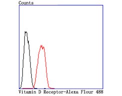
Flow cytometric analysis of Vitamin D Receptor was done on Hela cells. The cells were fixed, permeabilized and stained with Vitamin D Receptor antibody at 1/50 dilution (red) compared with an unlabelled control (cells without incubation with primary antibody; black). After incubation of the primary antibody on room temperature for an hour, the cells was stained with a Alexa Fluor 488-conjugated goat anti-rabbit IgG Secondary antibody at 1/500 dilution for 30 minutes.| Product Name | Vitamin D Receptor Recombinant Rabbit Monoclonal Antibody |
|---|---|
| Antibody Type | Primary Antibodies |
| Product description | This gene encodes the nuclear hormone receptor for vitamin D3. The active metabolite of vitamin D modulates the expression of a wide variety of genes in a developmentally specific manner. The receptor belongs to the family of trans-acting transcriptional regulatory factors and shows sequence similarity to the steroid and thyroid hormone receptors. This secosteroid hormone can up- or downregulate the expression of genes involved in a diverse array of responses such as proliferation, differentiation and calcium homeostasis. 1,25-(OH)2-vitamin D3 exerts its effects through interaction with the vitamin D receptor (VDR), a member of the superfamily of hormone-activated nuclear receptors. In its ligand-bound state, the VDR forms heterodimers with the 9-cis retinoic acid receptor, RXR, and affects gene expression by binding specific DNA sequences known as hormone response elements, or HREs. In addition to regulating the above-mentioned cellular responses, 1,25-(OH)2-vitamin D3 exhibits antiproliferative properties in osteosarcoma, melanoma, colon carcinoma and breast carcinoma cells. Mutations in this gene are associated with type II vitamin D-resistant rickets. A single nucleotide polymorphism in the initiation codon results in an alternate translation start site three codons downstream. Alternative splicing results in multiple transcript variants encoding different proteins. |
| Immunogen | Synthetic peptide with Human Vitamin D Receptor aa 1-50 / 427. |
| Clonality | Monoclonal |
|---|---|
| Isotype | IgG |
| Host Species | Recombinant rabbit |
| Tested Applications | FCIHCWB |
| WB:1:5000 IHC:1:50-1:200 FC:1:50-1:100 |
|
| Species Reactivity | HumanMouseRat |
| Concentration | 1mg/ml |
| Purification | Protein A |
| Alternative Names | 1 25 dihydroxyvitamin D3 receptor antibody
1 antibody
1 25 dihydroxyvitamin D3 receptor antibody 1 25-@dihydroxyvitamin D3 receptor antibody 25-dihydroxyvitamin D3 receptor antibody Member 1 antibody NR1I1 antibody Nuclear receptor subfamily 1 group I member 1 antibody PPP1R163 antibody Protein phosphatase 1 regulatory subunit 163 antibody VDR antibody VDR_HUMAN antibody Vitamin D (1 25- dihydroxyvitamin D3) receptor antibody Vitamin D hormone receptor antibody Vitamin D nuclear receptor variant 1 antibody Vitamin D receptor antibody Vitamin D3 receptor antibody |
|---|---|
| Molecular Weight(MW) | 48 kDa |
| Cellular Localization | Nucleus. |
WB
Western blot analysis of Vitamin D Receptor on different lysates with Rabbit anti-Vitamin D Receptor antibody at 1/5,000 dilution. Lane 1: HL-60 cell lysate (15 µg/Lane), Lane 2: T-47D cell lysate (15 µg/Lane), Lane 3: NIH/3T3 cell lysate (15 µg/Lane), Lane 4: L-929 cell lysate (15 µg/Lane), Lane 5: Rat kidney tissue lysate (20 µg/Lane), Exposure time: 30 seconds; 4-20% SDS-PAGE gel. Proteins were transferred to a PVDF membrane and blocked with 5% NFDM/TBST for 1 hour at room temperature. The primary antibody at 1/5,000 dilution was used in 5% NFDM/TBST at 4℃ overnight. Goat Anti-Rabbit IgG - HRP Secondary Antibody at 1/50,000 dilution was used for 1 hour at room temperature.
IHC
Immunohistochemical analysis of paraffin-embedded human skin tissue using anti-Vitamin D Receptor antibody. The section was pre-treated using heat mediated antigen retrieval with sodium citrate buffer (pH 6.0) for 20 minutes. The tissues were blocked in 5% BSA for 30 minutes at room temperature, washed with ddH2O and PBS, and then probed with the antibody (ET1704-09) at 1/100 dilution, for 30 minutes at room temperature and detected using an HRP conjugated compact polymer system. DAB was used as the chromogen. Counter stained with hematoxylin and mounted with DPX.
IHC
Immunohistochemical analysis of paraffin-embedded mouse colon tissue using anti-Vitamin D Receptor antibody. The section was pre-treated using heat mediated antigen retrieval with sodium citrate buffer (pH 6.0) for 20 minutes. The tissues were blocked in 5% BSA for 30 minutes at room temperature, washed with ddH2O and PBS, and then probed with the antibody (ET1704-09) at 1/100 dilution, for 30 minutes at room temperature and detected using an HRP conjugated compact polymer system. DAB was used as the chromogen. Counter stained with hematoxylin and mounted with DPX.
IHC
Immunohistochemical analysis of paraffin-embedded mouse skin tissue using anti-Vitamin D Receptor antibody. The section was pre-treated using heat mediated antigen retrieval with sodium citrate buffer (pH 6.0) for 20 minutes. The tissues were blocked in 5% BSA for 30 minutes at room temperature, washed with ddH2O and PBS, and then probed with the antibody (ET1704-09) at 1/100 dilution, for 30 minutes at room temperature and detected using an HRP conjugated compact polymer system. DAB was used as the chromogen. Counter stained with hematoxylin and mounted with DPX.
IHC
Immunohistochemical analysis of paraffin-embedded mouse colon tissue with Rabbit anti-Vitamin D Receptor antibody at 1/800 dilution. The section was pre-treated using heat mediated antigen retrieval with sodium citrate buffer (pH 6.0) (high pressure) for 2 minutes. The tissues were blocked in 1% BSA for 20 minutes at room temperature, washed with ddH2O and PBS, and then probed with the primary antibody at 1/800 dilution for 1 hour at room temperature. The detection was performed using an HRP conjugated compact polymer system. DAB was used as the chromogen. Tissues were counterstained with hematoxylin and mounted with DPX.
IHC
Immunohistochemical analysis of paraffin-embedded rat skin tissue with Rabbit anti-Vitamin D Receptor antibody at 1/800 dilution. The section was pre-treated using heat mediated antigen retrieval with sodium citrate buffer (pH 6.0) (high pressure) for 2 minutes. The tissues were blocked in 1% BSA for 20 minutes at room temperature, washed with ddH2O and PBS, and then probed with the primary antibody at 1/800 dilution for 1 hour at room temperature. The detection was performed using an HRP conjugated compact polymer system. DAB was used as the chromogen. Tissues were counterstained with hematoxylin and mounted with DPX.
FC
Flow cytometric analysis of Vitamin D Receptor was done on Hela cells. The cells were fixed, permeabilized and stained with Vitamin D Receptor antibody at 1/50 dilution (red) compared with an unlabelled control (cells without incubation with primary antibody; black). After incubation of the primary antibody on room temperature for an hour, the cells was stained with a Alexa Fluor 488-conjugated goat anti-rabbit IgG Secondary antibody at 1/500 dilution for 30 minutes.| Application Notes | WB:1:5000 IHC:1:50-1:200 FC:1:50-1:100 |
|---|
| Form | Liquid |
|---|---|
| Storage Instructions | Store at +4℃ after thawing. Aliquot store at -20℃. Avoid repeated freeze / thaw cycles. |
| Storage Buffer | 1*TBS (pH7.4), 1%BSA, 40%Glycerol. Preservative: 0.05% Sodium Azide. |


