Application
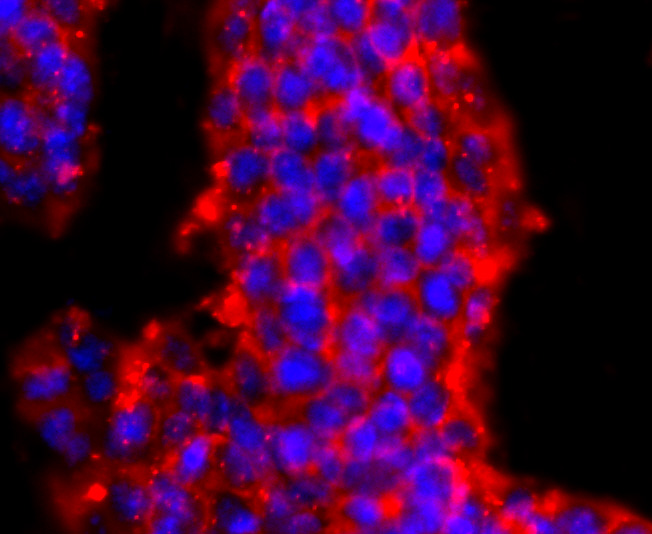
Fig1: ICC staining PRMT1 in D3 cells (red). The nuclear counter stain is DAPI (blue). Cells were fixed in paraformaldehyde, permeabilised with 0.25% Triton X100/PBS.Application
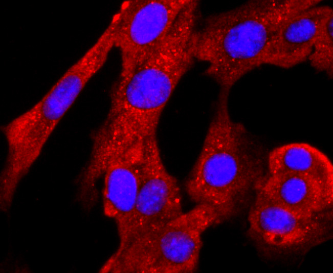
Fig2: ICC staining PRMT1 in NIH-3T3 cells (red). The nuclear counter stain is DAPI (blue). Cells were fixed in paraformaldehyde, permeabilised with 0.25% Triton X100/PBS.Application
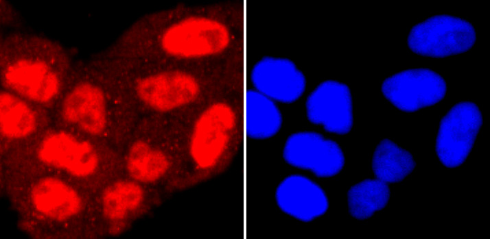
Fig3: ICC staining PRMT1 in Hela cells (red). The nuclear counter stain is DAPI (blue). Cells were fixed in paraformaldehyde, permeabilised with 0.25% Triton X100/PBS.Application
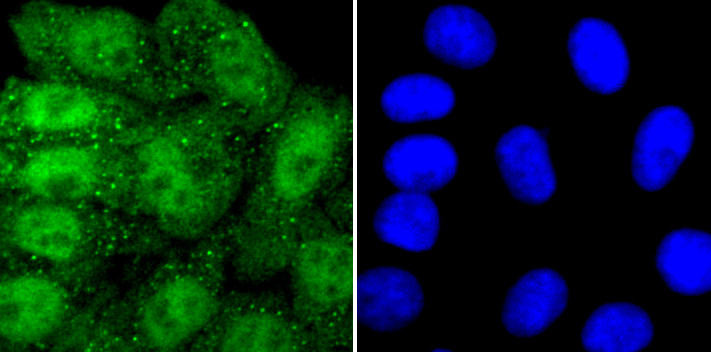
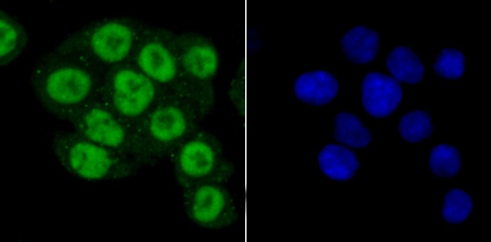
Fig4: ICC staining PRMT1 in HepG2 cells (green). The nuclear counter stain is DAPI (blue). Cells were fixed in paraformaldehyde, permeabilised with 0.25% Triton X100/PBS.Application
Fig5: ICC staining PRMT1 in SW480 cells (green). The nuclear counter stain is DAPI (blue). Cells were fixed in paraformaldehyde, permeabilised with 0.25% Triton X100/PBS.Application
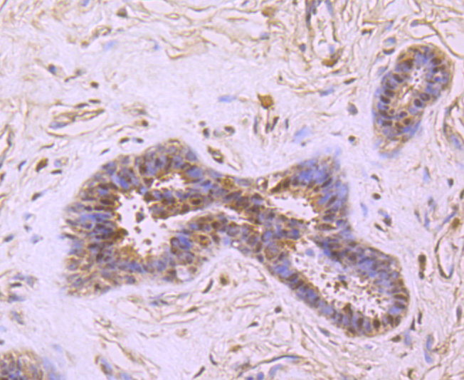
Fig6: Immunohistochemical analysis of paraffin-embedded human breast cancer tissue using anti-PRMT1 antibody. Counter stained with hematoxylin.Application
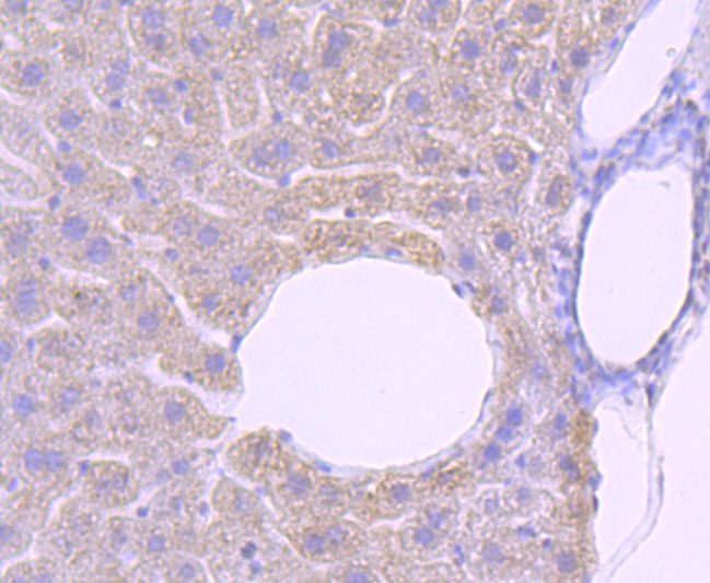
Fig7: Immunohistochemical analysis of paraffin-embedded mouse liver tissue using anti-PRMT1 antibody. Counter stained with hematoxylin.Application
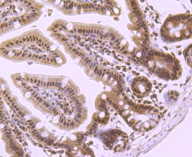
Fig8: Immunohistochemical analysis of paraffin-embedded mouse colon tissue using anti-PRMT1 antibody. Counter stained with hematoxylin.| Product Name | PRMT1 [JA11-17] |
|---|---|
| Antibody Type | Primary Antibodies |
| Product description | A class of proteins termed type 1 protein arginine N-methyltransferase (PRMT) enzymes contribute to posttranslational modification of RNA-binding proteins, but differ in substrate specificities, oligomerization properties and subcellular localization. PRMT1, the predominant form in mammalian cells, is located in the nucleus, while PRMT3 is present in the cytoplasm. At the carboxy-terminus, interleukin enhancer-binding factor 3 (ILF3) binds PRMT1, thereby regulating PRMT1 activitiy. Alternative mRNA splicing of the PRMT gene results in three isoforms of PRMT1 that differ in their amino-terminus regions. All three splice variants of PRMT1 are enzymatically active. PRMT3 recognizes and binds to RNA-associated substrates with a zinc-finger domain in its amino-terminus. The zinc-liganded form of this domain is required for the enzyme to recognize RNA-associated substrates. |
| Immunogen | recombinant protein |
| Clonality | Monoclonal |
|---|---|
| Isotype | IgG |
| Host Species | Recombinant rabbit |
| Tested Applications | WB,ICC/IF,IHC,ChIP |
| WB:1:500-1:1,000 ICC:1:100-1:500 IHC:1:50-1:200 |
|
| Species Reactivity | Human,Mouse,Rat |
| Concentration | 1mg/ml |
| Purification | Unpurified |
| Alternative Names | ANM 1 antibody ANM1 antibody ANM1_HUMAN antibody HCP 1 antibody HCP1 antibody Heterogeneous nuclear ribonucleoprotein methyltransferase 1 like 2 antibody Heterogeneous nuclear ribonucleoproteins methyltransferase like 2 antibody Heterogeneous nuclear ribonucleoproteins methyltransferase like2 antibody Histone-arginine N-methyltransferase PRMT1 antibody HMT 2 antibody HMT1 (hnRNP methyltransferase S. cerevisiae) like 2 antibody HMT1 hnRNP methyltransferase antibody HMT1 hnRNP methyltransferase like 2 (S. cerevisiae) antibody HMT1 hnRNP methyltransferase like 2 antibody HMT2 antibody HRMT1 L2 antibody HRMT1L 2 antibody HRMT1L2 antibody Human mRNA for suppressor for yeast mutant antibody Human mRNA for suppressor for yeast mutant complete cds antibody Interferon receptor 1 bound protein 4 antibody Interferon receptor 1 bound protein4 antibody Interferon receptor 1-bound protein 4 antibody Interferon receptor 1bound protein 4 antibody IR1 B4 antibody IR1B 4 antibody IR1B4 antibody Mrmt 1 antibody Mrmt1 antibody PRMT 1 antibody PRMT1 antibody Protein arginine methyltransferase 1 antibody Protein arginine N methyltransferase 1 antibody Protein arginine N methyltransferase1 antibody Protein arginine N-methyltransferase 1 antibody R1B4 antibody S. cerevisiae like 2 antibody |
|---|---|
| Molecular Weight(MW) | 42 kDa |
| Cellular Localization | Nucleus. Cytoplasm. |
Application
Fig1: ICC staining PRMT1 in D3 cells (red). The nuclear counter stain is DAPI (blue). Cells were fixed in paraformaldehyde, permeabilised with 0.25% Triton X100/PBS.
Application
Fig2: ICC staining PRMT1 in NIH-3T3 cells (red). The nuclear counter stain is DAPI (blue). Cells were fixed in paraformaldehyde, permeabilised with 0.25% Triton X100/PBS.
Application
Fig3: ICC staining PRMT1 in Hela cells (red). The nuclear counter stain is DAPI (blue). Cells were fixed in paraformaldehyde, permeabilised with 0.25% Triton X100/PBS.
Application
Fig4: ICC staining PRMT1 in HepG2 cells (green). The nuclear counter stain is DAPI (blue). Cells were fixed in paraformaldehyde, permeabilised with 0.25% Triton X100/PBS.
Application
Fig5: ICC staining PRMT1 in SW480 cells (green). The nuclear counter stain is DAPI (blue). Cells were fixed in paraformaldehyde, permeabilised with 0.25% Triton X100/PBS.
Application
Fig6: Immunohistochemical analysis of paraffin-embedded human breast cancer tissue using anti-PRMT1 antibody. Counter stained with hematoxylin.
Application
Fig7: Immunohistochemical analysis of paraffin-embedded mouse liver tissue using anti-PRMT1 antibody. Counter stained with hematoxylin.
Application
Fig8: Immunohistochemical analysis of paraffin-embedded mouse colon tissue using anti-PRMT1 antibody. Counter stained with hematoxylin.| Positive Control | D3, HepG2, NIH-3T3, Hela, SW480, human breast cancer tissue, mouse liver tissue, mouse colon tissue. |
|---|---|
| Application Notes | WB:1:500-1:1,000 ICC:1:100-1:500 IHC:1:50-1:200 |
| Form | Liquid |
|---|---|
| Storage Instructions | Store at +4℃ after thawing. Aliquot store at -20℃ or -80℃. Avoid repeated freeze / thaw cycles. |
| Storage Buffer | 1*TBS (pH7.4), 1%BSA, 40%Glycerol. Preservative: 0.05% Sodium Azide. |


