Application
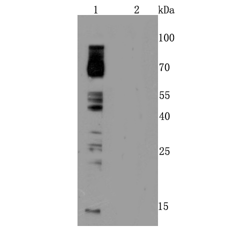
Fig1: Western blot analysis of Phosphotyrosine on A431 cells lysates treated ( Line 1) and untreated with Sodium metavanadate using anti- Phosphotyrosine antibody at 1/100 dilution.Application
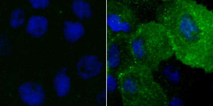
Fig2: ICC staining Phosphotyrosine in A431 cells (green) nontreated (Left) or treated with Sodium metavanadate (Right). The nuclear counter stain is DAPI (blue). Cells were fixed in paraformaldehyde, permeabilised with 0.25% Triton X100/PBS.Application
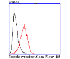
Fig3: Flow cytometric analysis of A431 cells with Phosphotyrosine antibody at 1/50 dilution (red) compared with an unlabelled control (cells without incubation with primary antibody; black). Alexa Fluor 488-conjugated goat anti rabbit IgG was used as the secondary antibody.| Product Name | Phosphotyrosine Recombinant Rabbit Monoclonal Antibody |
|---|---|
| Antibody Type | Primary Antibodies |
| Product description | Tyrosine phosphorylation plays a key role in cellular signaling. Research studies have shown that in cancer, unregulated tyrosine kinase activity can drive malignancy and tumor formation by generating inappropriate proliferation and survival signals. Antibodies specific for phospho-tyrosine have been invaluable reagents in these studies. The phospho-tyrosine monoclonal antibodies developed by Cell Signaling Technology are exceptionally sensitive tools for studying tyrosine phosphorylation and monitoring tyrosine kinase activity in high throughput drug discovery. |
| Immunogen | Synthetic peptide conjugated to Keyhole Limpet Haemocyanin (KLH) |
| Clonality | monoclonal |
|---|---|
| Isotype | IgG |
| Host Species | Recombinant rabbit |
| Tested Applications | FCICC/IFWB |
| WB:1:50-1:200 ICC/IF:1:50 FC:1:10-1:50 |
|
| Species Reactivity | HumanMouseRat |
| Concentration | 1mg/ml |
| Purification | Protein A |
Application
Fig1: Western blot analysis of Phosphotyrosine on A431 cells lysates treated ( Line 1) and untreated with Sodium metavanadate using anti- Phosphotyrosine antibody at 1/100 dilution.
Application
Fig2: ICC staining Phosphotyrosine in A431 cells (green) nontreated (Left) or treated with Sodium metavanadate (Right). The nuclear counter stain is DAPI (blue). Cells were fixed in paraformaldehyde, permeabilised with 0.25% Triton X100/PBS.
Application
Fig3: Flow cytometric analysis of A431 cells with Phosphotyrosine antibody at 1/50 dilution (red) compared with an unlabelled control (cells without incubation with primary antibody; black). Alexa Fluor 488-conjugated goat anti rabbit IgG was used as the secondary antibody.| Positive Control | A431 cells lysates treated with Sodium metavanadate. |
|---|---|
| Application Notes | WB:1:50-1:200 ICC/IF:1:50 FC:1:10-1:50 |
| Form | Liquid |
|---|---|
| Storage Instructions | Store at +4℃ after thawing. Aliquot store at -20℃ or -80℃. Avoid repeated freeze / thaw cycles. |
| Storage Buffer | 1*TBS (pH7.4), 1%BSA, 40%Glycerol. Preservative: 0.05% Sodium Azide. |


