WB
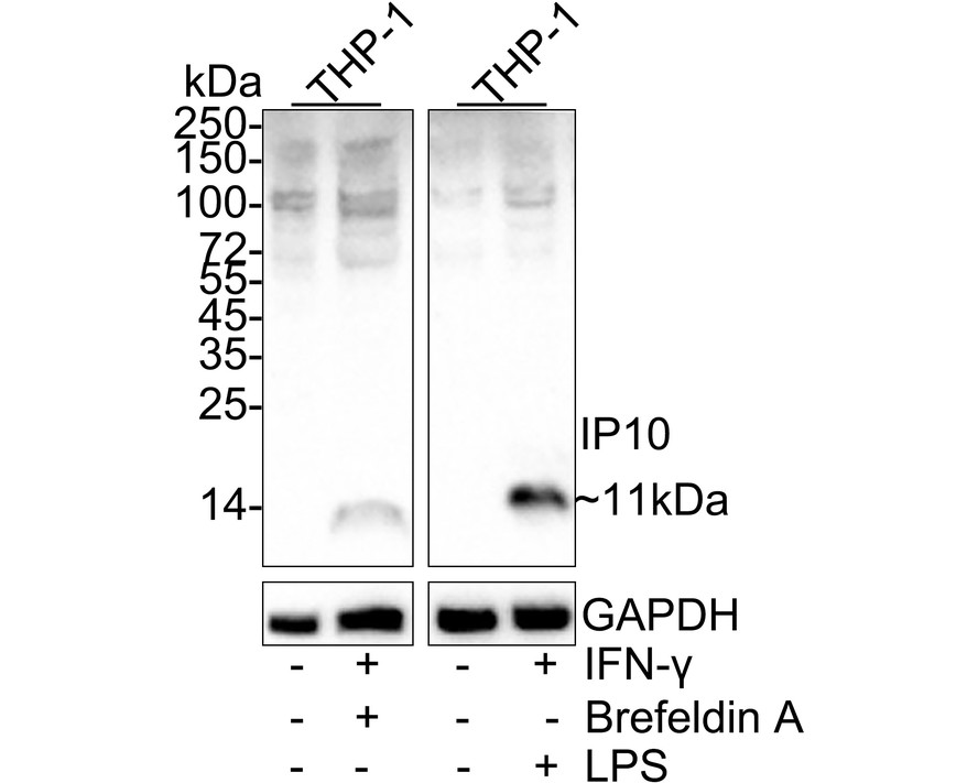
Western blot analysis of IP10 on different lysates with Rabbit anti-IP10 antibody at 1/1,000 dilution. Lane 1: THP-1 cell lysate, Lane 2: THP-1 treated with 100ng/mL IFN-γ for 24 hours then 300ng/mL Brefeldin A for 20 hours cell lysate, Lane 3: THP-1 cell lysate, Lane 4: THP-1 treated with 200ng/mL IFN-γ and 50ng/mL LPS for 24 hours cell lysate, Lysates/proteins at 20 µg/Lane. Exposure time: 3 minutes; 4-20% SDS-PAGE gel. Proteins were transferred to a PVDF membrane and blocked with 5% NFDM/TBST for 1 hour at room temperature. The primary antibody at 1/1,000 dilution was used in 5% NFDM/TBST at 4℃ overnight. Goat Anti-Rabbit IgG - HRP Secondary Antibody at 1/50,000 dilution was used for 1 hour at room temperature.IHC
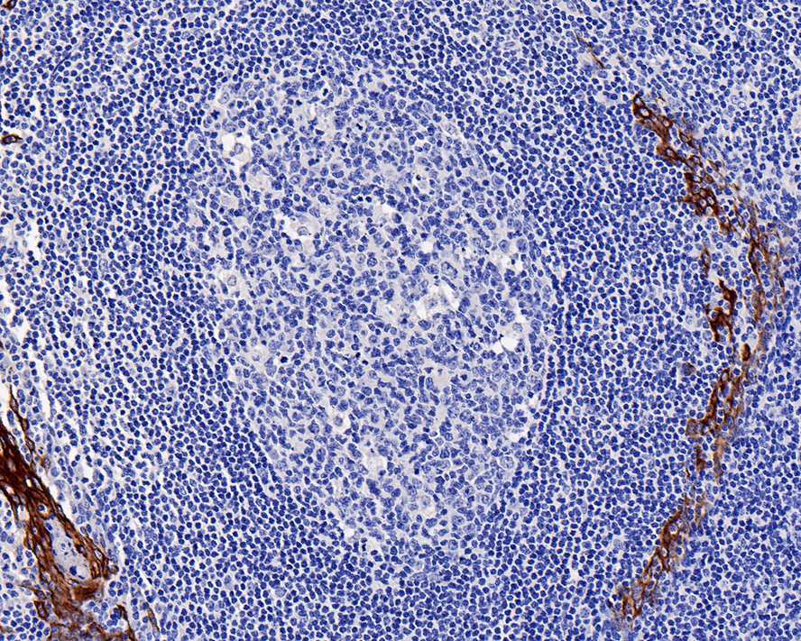
Immunohistochemical analysis of paraffin-embedded human tonsil tissue with Rabbit anti-IP10 antibody at 1/1,000 dilution. The section was pre-treated using heat mediated antigen retrieval with Tris-EDTA buffer (pH 9.0) for 20 minutes. The tissues were blocked in 1% BSA for 20 minutes at room temperature, washed with ddH2O and PBS, and then probed with the primary antibody at 1/1,000 dilution for 1 hour at room temperature. The detection was performed using an HRP conjugated compact polymer system. DAB was used as the chromogen. Tissues were counterstained with hematoxylin and mounted with DPX.ICC/IF
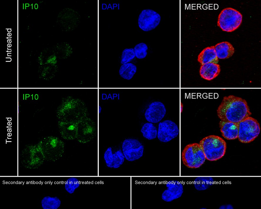
Immunocytochemistry analysis of THP-1 cells treated with 200ng/mL IFN-γ then treated with 50ng/mL LPS for 24 hours labeling IP10 with Rabbit anti-IP10 antibody at 1/100 dilution. Cells were fixed in 4% paraformaldehyde for 20 minutes at room temperature, permeabilized with 0.1% Triton X-100 in PBS for 5 minutes at room temperature, then blocked with 1% BSA in 10% negative goat serum for 1 hour at room temperature. Cells were then incubated with Rabbit anti-IP10 antibody at 1/100 dilution in 1% BSA in PBST overnight at 4 ℃. Goat Anti-Rabbit IgG H&L (488) was used as the secondary antibody at 1/1,000 dilution. PBS instead of the primary antibody was used as the secondary antibody only control. Nuclear DNA was labelled in blue with DAPI.IF-P
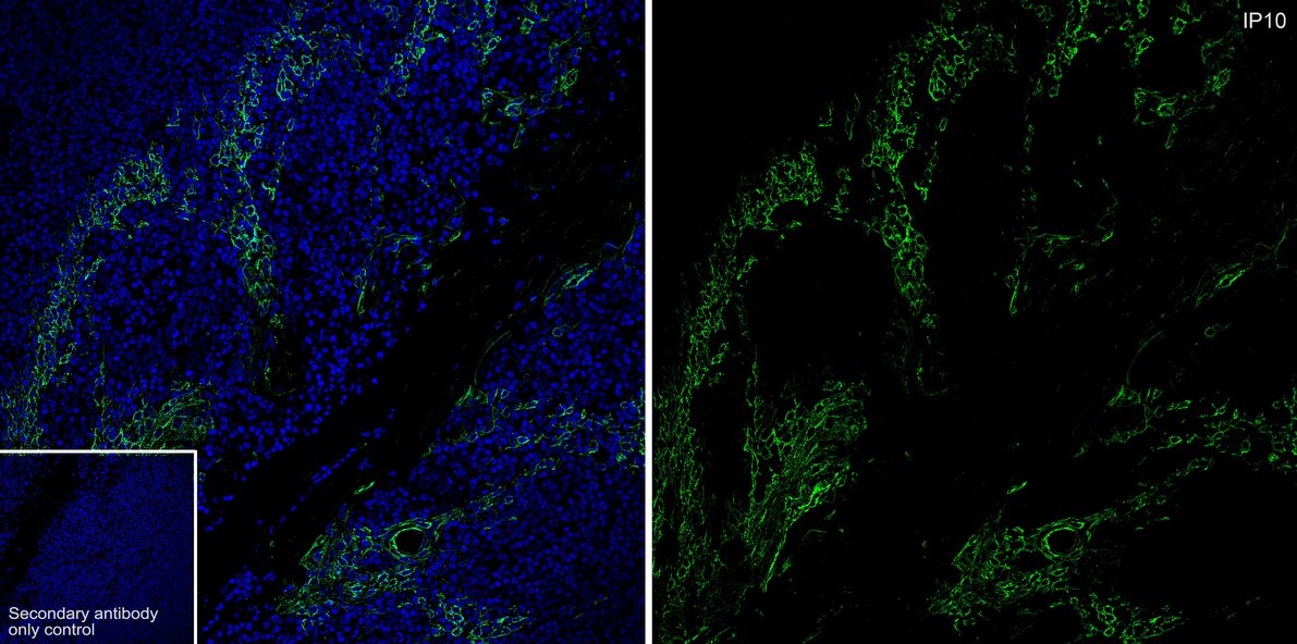
Species: Human, Site: tonsil, Sample: Paraffin-embedded section, Antibody concentration: 1/500.| Product Name | IP10 Recombinant Rabbit Monoclonal Antibody |
|---|---|
| Antibody Type | Primary Antibodies |
| Product description | Chemokines are members of a superfamily of inducible, secreted, pro-inflammatory cytokines. Members of the chemokine family exhibit 20% to 50% homology in their predicted amino acid sequences and are divided into four subfamilies: C-C, C-X-C, C and C-X3-C. In the C-X-C or α subfamily, the first two of four cysteine motifs are separated by another amino acid residue. In the second subfamily, designated C-C or β, the first cysteines are adjacent. C subfamily members, also designated γ chemokines, lack the first and third cysteine residues of the conserved motif. In the C-X3-C, or δ subfamily, members have three amino acids between the two cysteines. The C-X-C chemokine subfamily includes IL-8, GROα/β/γ (and the murine homologs KC, MIP-2α and MIP-2β), platelet basic protein, ENA-78, GCP-2, PF4, IP-10 (and its murine homolog, CRG) and MIG. |
| Immunogen | Synthetic peptide within Human IP10 aa 49-98 / 98. |
| Clonality | monoclonal |
|---|---|
| Isotype | IgG |
| Host Species | Recombinant rabbit |
| Tested Applications | ICC/IFIF-PIHCWB |
| WB:1:1000 IHC:1:50-1:1000 ICC/IF:1:50-1:100 IF-P:1:500 |
|
| Species Reactivity | Human |
| Concentration | 1mg/ml |
| Purification | Protein A |
| Alternative Names | Interferon gamma induced factor MOB1 mouse homolog of antibody Interferon gamma induced protein 10 antibody 10 kDa interferon gamma induced protein antibody 10 kDa interferon gamma-induced protein antibody C X C motif chemokine 10 antibody C7 antibody Chemokine (C X C motif) ligand 10 antibody Chemokine CXC motif ligand 10 antibody Crg 2 antibody CRG2 antibody CXCL10 antibody CXCL10(1-73) antibody CXL10_HUMAN antibody Gamma IP10 antibody Gamma-IP10 antibody gIP 10 antibody GIP10 antibody IFI10 antibody INP 10 antibody INP10 antibody Interferon activated gene 10 antibody Interferon activated gene 10 antibody Interferon gamma induced cell line antibody Interferon inducible cytokine IP 10 antibody Interferon inducible cytokine IP10 antibody IP 10 antibody IP-10 antibody Mob 1 antibody MOB1 antibody Protein 10 from interferon (gamma) induced cell line antibody SCYB10 antibody Small inducible cytokine B10 antibody Small inducible cytokine B10 precursor antibody Small inducible cytokine subfamily B (Cys X Cys) member 10 antibody Small inducible cytokine subfamily B CXC member 10 antibody Small inducible cytokine subfamily B member 10 antibody Small-inducible cytokine B10 antibody |
|---|---|
| Molecular Weight(MW) | 11kDa |
| Cellular Localization | Secreted. |
| SwissProt ID | P02778 |
|---|
WB
Western blot analysis of IP10 on different lysates with Rabbit anti-IP10 antibody at 1/1,000 dilution. Lane 1: THP-1 cell lysate, Lane 2: THP-1 treated with 100ng/mL IFN-γ for 24 hours then 300ng/mL Brefeldin A for 20 hours cell lysate, Lane 3: THP-1 cell lysate, Lane 4: THP-1 treated with 200ng/mL IFN-γ and 50ng/mL LPS for 24 hours cell lysate, Lysates/proteins at 20 µg/Lane. Exposure time: 3 minutes; 4-20% SDS-PAGE gel. Proteins were transferred to a PVDF membrane and blocked with 5% NFDM/TBST for 1 hour at room temperature. The primary antibody at 1/1,000 dilution was used in 5% NFDM/TBST at 4℃ overnight. Goat Anti-Rabbit IgG - HRP Secondary Antibody at 1/50,000 dilution was used for 1 hour at room temperature.
IHC
Immunohistochemical analysis of paraffin-embedded human tonsil tissue with Rabbit anti-IP10 antibody at 1/1,000 dilution. The section was pre-treated using heat mediated antigen retrieval with Tris-EDTA buffer (pH 9.0) for 20 minutes. The tissues were blocked in 1% BSA for 20 minutes at room temperature, washed with ddH2O and PBS, and then probed with the primary antibody at 1/1,000 dilution for 1 hour at room temperature. The detection was performed using an HRP conjugated compact polymer system. DAB was used as the chromogen. Tissues were counterstained with hematoxylin and mounted with DPX.
ICC/IF
Immunocytochemistry analysis of THP-1 cells treated with 200ng/mL IFN-γ then treated with 50ng/mL LPS for 24 hours labeling IP10 with Rabbit anti-IP10 antibody at 1/100 dilution. Cells were fixed in 4% paraformaldehyde for 20 minutes at room temperature, permeabilized with 0.1% Triton X-100 in PBS for 5 minutes at room temperature, then blocked with 1% BSA in 10% negative goat serum for 1 hour at room temperature. Cells were then incubated with Rabbit anti-IP10 antibody at 1/100 dilution in 1% BSA in PBST overnight at 4 ℃. Goat Anti-Rabbit IgG H&L (488) was used as the secondary antibody at 1/1,000 dilution. PBS instead of the primary antibody was used as the secondary antibody only control. Nuclear DNA was labelled in blue with DAPI.
IF-P
Species: Human, Site: tonsil, Sample: Paraffin-embedded section, Antibody concentration: 1/500.| Application Notes | WB:1:1000 IHC:1:50-1:1000 ICC/IF:1:50-1:100 IF-P:1:500 |
|---|
| Form | Liquid |
|---|---|
| Storage Instructions | Store at +4℃ after thawing. Aliquot store at -20℃ or -80℃. Avoid repeated freeze / thaw cycles. |
| Storage Buffer | 1*TBS (pH7.4), 1%BSA, 40%Glycerol. Preservative: 0.05% Sodium Azide. |


