WB
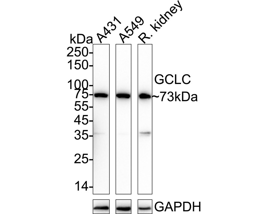
Western blot analysis of GCLC on different lysates with Rabbit anti-GCLC antibody at 1/1,000 dilution. Lane 1: A431 cell lysate (20 µg/Lane), Lane 2: A549 cell lysate (20 µg/Lane), Lane 3: Rat kidney tissue lysate (40 µg/Lane), Exposure time: 1 minute; 4-20% SDS-PAGE gel. Proteins were transferred to a PVDF membrane and blocked with 5% NFDM/TBST for 1 hour at room temperature. The primary antibody at 1/1,000 dilution was used in 5% NFDM/TBST at 4℃ overnight. Goat Anti-Rabbit IgG - HRP Secondary Antibody at 1/50,000 dilution was used for 1 hour at room temperature.IHC
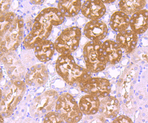
Immunohistochemical analysis of paraffin-embedded rat kidney tissue using anti-GCLC antibody. Counter stained with hematoxylin.IHC
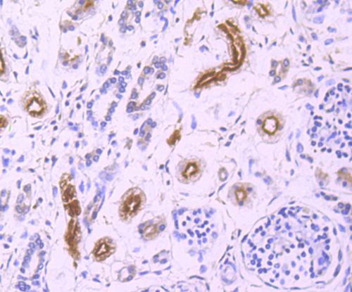
Immunohistochemical analysis of paraffin-embedded human fetal kidney tissue using anti-GCLC antibody. Counter stained with hematoxylin.IHC
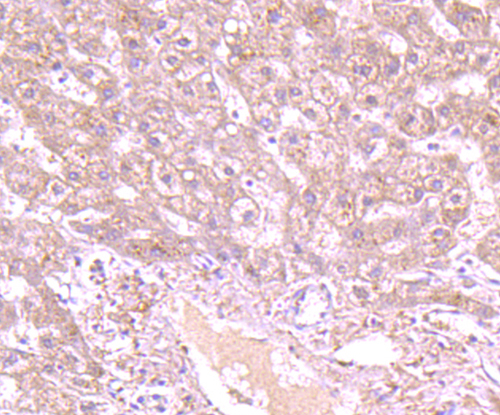
Immunohistochemical analysis of paraffin-embedded human fetal liver tissue using anti-GCLC antibody. Counter stained with hematoxylin.FC
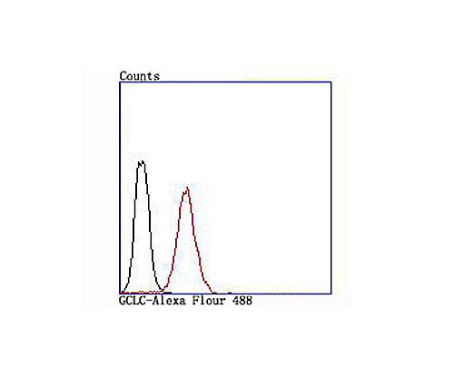
Flow cytometric analysis of Jurkat cells with GCLC antibody at 1/100 dilution (red) compared with an unlabelled control (cells without incubation with primary antibody; black).| Product Name | GCLC Recombinant Rabbit Monoclonal Antibody |
|---|---|
| Antibody Type | Primary Antibodies |
| Product description | The GCLC gene consists of 16 exons and encodes the 636 amino acid protein g-GCSc (g-glutamylcysteine synthetase heavy subunit), also designated g-L-glutamate-L-cysteine ligase catalytic subunit (GLCLC). g-GCSc is expressed in hemocytes, brain, liver and kidney. g-GCSc associates with a regulatory or modifier subunit, g-GCSm (g-glutamylcysteine synthetase light subunit), to form a heterodimer, g-GCS. g-GCS is the first enzyme involved and the rate determining step in glutathione biosynthesis. Oxidants, cadium and methyl mercury upregulate the transcription of g-GCS. H2O2 regulation depends on the Yap1 protein and the presence of glutamate, glutamine and lysine. Cadium regulates transcription through proteins Met-4, Met-31 and Met-32. Cbf1, a DNA binding protein, inhibits transcription of g-GCS. Chemopreventive compounds cause increased levels of g-GCSc in kidney tissues, which may protect against chemically induced carcinogenesis. A His370Leu amino acid change in g-GCSc causes deficiencies in activity which are responsible for hemolytic anemia and low red blood cell glutathione levels. |
| Immunogen | Synthetic peptide within Human GCLC aa 588-637 / 637. |
| Clonality | Monoclonal |
|---|---|
| Isotype | IgG |
| Host Species | Recombinant rabbit |
| Tested Applications | FCIHCWB |
| WB:1:500-1:1000 IHC:1:501:200 FC:1:50-1:100 |
|
| Species Reactivity | HumanRat |
| Concentration | 1mg/ml |
| Purification | Protein A |
| Alternative Names | EC 6.3.2.2 antibody Gamma ECS antibody Gamma glutamylcysteine synthetase antibody Gamma-ECS antibod Gamma-glutamylcysteine synthetase antibody GCL antibody Gclc antibody GCS antibody GCS heavy chain antibody GLCL antibody GLCLC antibody Glutamate cysteine ligase catalytic subunit antibody Glutamatecysteine ligase catalytic subunit antibody GSH1_HUMAN antibody |
|---|---|
| Molecular Weight(MW) | 73 kDa |
| Cellular Localization | Cytoplasm. |
WB
Western blot analysis of GCLC on different lysates with Rabbit anti-GCLC antibody at 1/1,000 dilution. Lane 1: A431 cell lysate (20 µg/Lane), Lane 2: A549 cell lysate (20 µg/Lane), Lane 3: Rat kidney tissue lysate (40 µg/Lane), Exposure time: 1 minute; 4-20% SDS-PAGE gel. Proteins were transferred to a PVDF membrane and blocked with 5% NFDM/TBST for 1 hour at room temperature. The primary antibody at 1/1,000 dilution was used in 5% NFDM/TBST at 4℃ overnight. Goat Anti-Rabbit IgG - HRP Secondary Antibody at 1/50,000 dilution was used for 1 hour at room temperature.
IHC
Immunohistochemical analysis of paraffin-embedded rat kidney tissue using anti-GCLC antibody. Counter stained with hematoxylin.
IHC
Immunohistochemical analysis of paraffin-embedded human fetal kidney tissue using anti-GCLC antibody. Counter stained with hematoxylin.
IHC
Immunohistochemical analysis of paraffin-embedded human fetal liver tissue using anti-GCLC antibody. Counter stained with hematoxylin.
FC
Flow cytometric analysis of Jurkat cells with GCLC antibody at 1/100 dilution (red) compared with an unlabelled control (cells without incubation with primary antibody; black).| Positive Control | A431, rat kidney tissue, human fetal kidney tissue, human fetal liver tissue, Jurkat. |
|---|---|
| Application Notes | WB:1:500-1:1000 IHC:1:501:200 FC:1:50-1:100 |
| Form | Liquid |
|---|---|
| Storage Instructions | Store at +4℃ after thawing. Aliquot store at -20℃ or -80℃. Avoid repeated freeze / thaw cycles. |
| Storage Buffer | 1*TBS (pH7.4), 1%BSA, 40%Glycerol. Preservative: 0.05% Sodium Azide. |


