WB
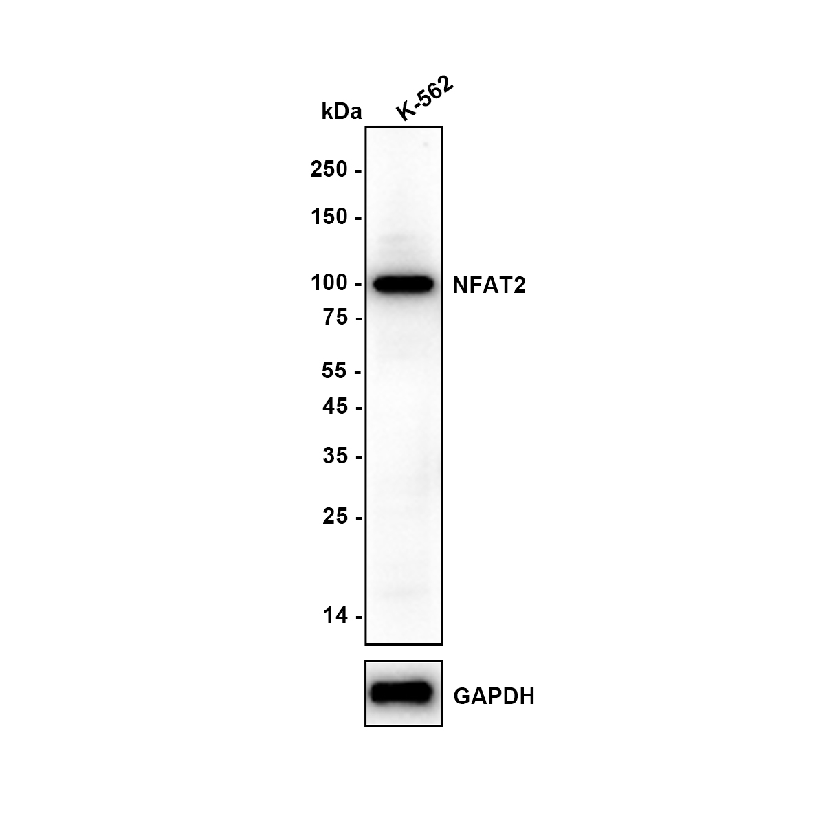
Western blot analysis of NFAT2 on K-562 (Human chronic myelogenous leukemia cell) lysate with Rabbit anti-NFAT2 antibody at 1/1,000 dilution. Lysates/proteins at 10 µg/Lane. Exposure time: 59 seconds; Blocking: 5% NFDM/TBST, 1 hour at room temperature, Primary antibody: 1/1,000 in 5% NFDM/TBST, overnight at 4 ℃, Secondary antibody: Goat anti-Rabbit IgG-HRP, 1/50,000 in 5% NFDM/TBST, 1 hour at room temperature.IHC
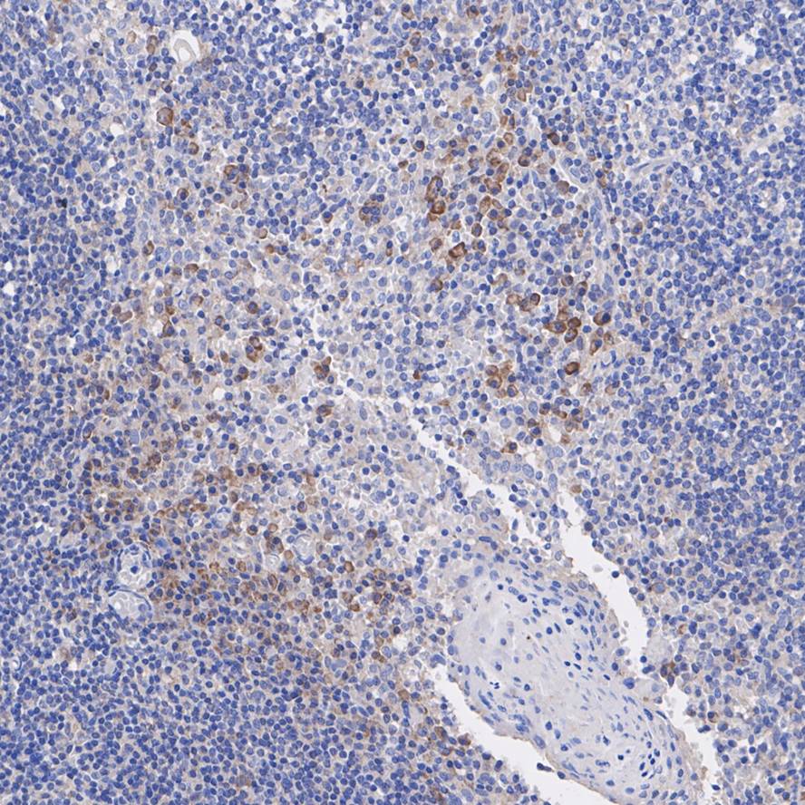
Immunohistochemical analysis of paraffin-embedded human tonsil tissue with Rabbit anti-NFAT2 antibody at 1/200 dilution. The section was pre-treated using heat mediated antigen retrieval with sodium citrate buffer (pH 6.0) (high pressure) for 2 minutes. The tissues were blocked in 1% BSA for 20 minutes at room temperature, washed with ddH2O and PBS, and then probed with the primary antibody at 1/200 dilution for 1 hour at room temperature. The detection was performed using an HRP conjugated compact polymer system. DAB was used as the chromogen. Tissues were counterstained with hematoxylin and mounted with DPX.FC
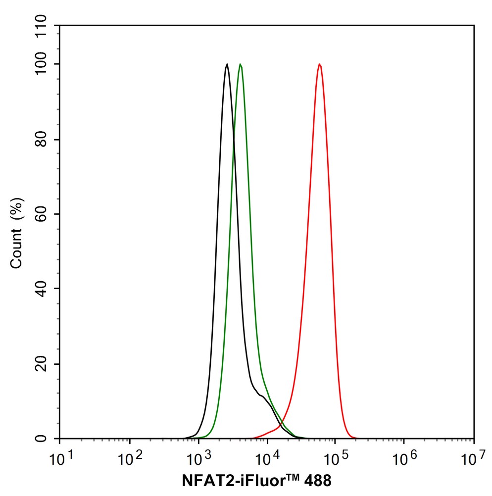
Flow cytometric analysis of K-562 cells labeling NFAT2. Cells were fixed and permeabilized. Then stained with the primary antibody (1/1,000) (red) compared with Rabbit IgG Isotype Control (green). After incubation of the primary antibody at +4℃ for an hour, the cells were stained with a 488 conjugate-Goat anti-Rabbit IgG Secondary antibody at 1/1,000 dilution for 30 minutes at +4℃. Unlabelled sample was used as a control (cells without incubation with primary antibody; black).| Product Name | NFAT2 Recombinant Rabbit Monoclonal Antibody |
|---|---|
| Antibody Type | Primary Antibodies |
| Product description | Plays a role in the inducible expression of cytokine genes in T-cells, especially in the induction of the IL-2 or IL-4 gene transcription. Also controls gene expression in embryonic cardiac cells. Could regulate not only the activation and proliferation but also the differentiation and programmed death of T-lymphocytes as well as lymphoid and non-lymphoid cells. Required for osteoclastogenesis and regulates many genes important for osteoclast differentiation and function. |
| Immunogen | Synthetic peptide within Human NFAT2 aa 894-943 / 943. |
| Clonality | Monoclonal |
|---|---|
| Isotype | IgG |
| Host Species | Recombinant rabbit |
| Tested Applications | FCIHCWB |
| WB:1:500-1:1000 IHC:1:50-1:200 FC:1:1000 |
|
| Species Reactivity | Human |
| Concentration | 1mg/ml |
| Purification | Protein A |
| Alternative Names | cytoplasmic 1 antibody MGC138448 antibody NF ATc antibody NF ATc1 antibody NF-ATc antibody NF-ATc1 antibody NF-ATc1.2 antibody NFAC1_HUMAN antibody NFAT 2 antibody NFAT transcription complex cytosolic component antibody NFATC 1 antibody NFATc antibody NFATc1 antibody Nuclear factor of activated T cells cytoplasmic 1 antibody Nuclear factor of activated T cells cytoplasmic calcineurin dependent 1 antibody Nuclear factor of activated T cells cytosolic component 1 antibody nuclear factor of activated T-cells 'c' antibody Nuclear factor of activated T-cells antibody |
|---|---|
| Molecular Weight(MW) | 101 kDa |
| Cellular Localization | Cytoplasm. Nucleus. |
| SwissProt ID | O95644 |
|---|
WB
Western blot analysis of NFAT2 on K-562 (Human chronic myelogenous leukemia cell) lysate with Rabbit anti-NFAT2 antibody at 1/1,000 dilution. Lysates/proteins at 10 µg/Lane. Exposure time: 59 seconds; Blocking: 5% NFDM/TBST, 1 hour at room temperature, Primary antibody: 1/1,000 in 5% NFDM/TBST, overnight at 4 ℃, Secondary antibody: Goat anti-Rabbit IgG-HRP, 1/50,000 in 5% NFDM/TBST, 1 hour at room temperature.
IHC
Immunohistochemical analysis of paraffin-embedded human tonsil tissue with Rabbit anti-NFAT2 antibody at 1/200 dilution. The section was pre-treated using heat mediated antigen retrieval with sodium citrate buffer (pH 6.0) (high pressure) for 2 minutes. The tissues were blocked in 1% BSA for 20 minutes at room temperature, washed with ddH2O and PBS, and then probed with the primary antibody at 1/200 dilution for 1 hour at room temperature. The detection was performed using an HRP conjugated compact polymer system. DAB was used as the chromogen. Tissues were counterstained with hematoxylin and mounted with DPX.
FC
Flow cytometric analysis of K-562 cells labeling NFAT2. Cells were fixed and permeabilized. Then stained with the primary antibody (1/1,000) (red) compared with Rabbit IgG Isotype Control (green). After incubation of the primary antibody at +4℃ for an hour, the cells were stained with a 488 conjugate-Goat anti-Rabbit IgG Secondary antibody at 1/1,000 dilution for 30 minutes at +4℃. Unlabelled sample was used as a control (cells without incubation with primary antibody; black).| Application Notes | WB:1:500-1:1000 IHC:1:50-1:200 FC:1:1000 |
|---|
| Form | Liquid |
|---|---|
| Storage Instructions | Store at +4℃ after thawing. Aliquot store at -20℃ or -80℃. Avoid repeated freeze / thaw cycles. |
| Storage Buffer | 1*TBS (pH7.4), 1%BSA, 40%Glycerol. Preservative: 0.05% Sodium Azide. |


