Application
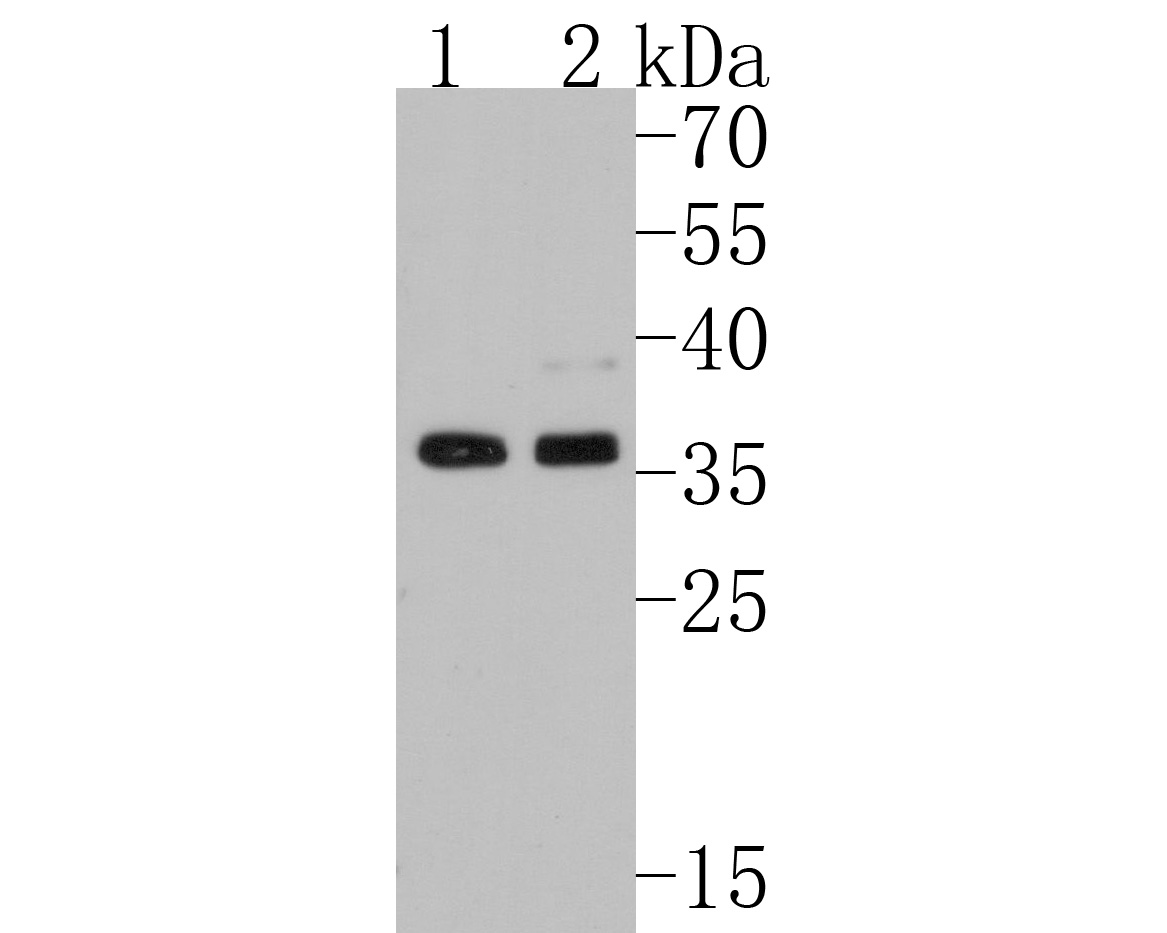
Fig1: Western blot analysis of hnRNP A1 on different lysates. Proteins were transferred to a PVDF membrane and blocked with 5% BSA in PBS for 1 hour at room temperature. The primary antibody (1/500) was used in 5% BSA at room temperature for 2 hours. Goat Anti-Rabbit IgG - HRP Secondary Antibody at 1:200,000 dilution was used for 1 hour at room temperature. Positive control: Lane 1: HepG2 cell lysate, Lane 2: PC-12 cell lysate.Application
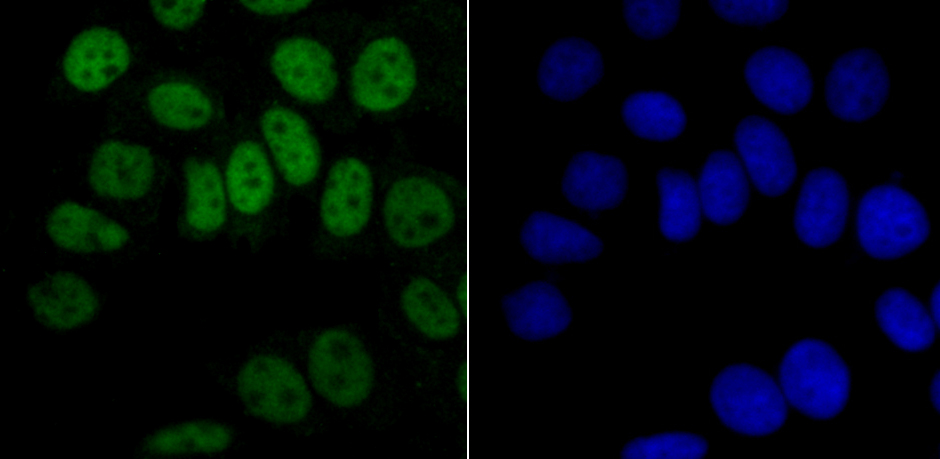
Fig2: ICC staining hnRNP A1 in MCF-7 cells (green). The nuclear counter stain is DAPI (blue). Cells were fixed in paraformaldehyde, permeabilised with 0.25% Triton X100/PBS.Application
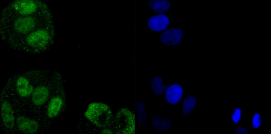
Fig3: ICC staining hnRNP A1 in Hela cells (green). The nuclear counter stain is DAPI (blue). Cells were fixed in paraformaldehyde, permeabilised with 0.25% Triton X100/PBS.Application
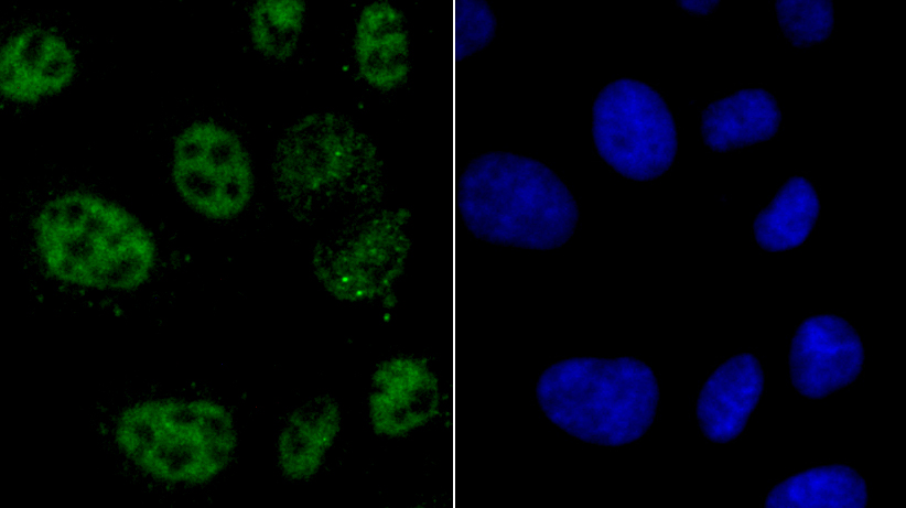
Fig4: ICC staining hnRNP A1 in HepG2 cells (green). The nuclear counter stain is DAPI (blue). Cells were fixed in paraformaldehyde, permeabilised with 0.25% Triton X100/PBS.Application
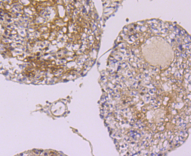
Fig5: Immunohistochemical analysis of paraffin-embedded rat testis tissue using anti-hnRNP A1 antibody. Counter stained with hematoxylin.Application
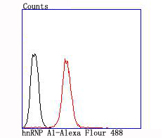
Fig6: Flow cytometric analysis of Jurkat cells with hnRNP A1 antibody at 1/100 dilution (red) compared with an unlabelled control (cells without incubation with primary antibody; black).| Product Name | hnRNP A1 Recombinant Rabbit Monoclonal Antibody |
|---|---|
| Antibody Type | Primary Antibodies |
| Product description | Heterogeneous nuclear ribonucleoproteins (hnRNPs) constitute a set of poly-peptides that contribute to mRNA transcription and pre-mRNA processing as well as mature mRNA transport to the cytoplasm and translation. They also bind heterogeneous nuclear RNA (hnRNA), which are the transcripts produced by RNA polymerase II. There are approximately 20 known hnRNP proteins, and their complexes are the major constituents of the spliceosome. The majority of hnRNP protein components are localized to the nucleus; however some shuttle between the nucleus and the cytoplasm. The A/B subfamily of hnRNPs include A1, A2/B1, A3 and A0, and in Xenopus, hnRNP A1, A2 and A3 are ubiquitously expressed throughout development as well as in adult tissues. hnRNP A1 and A2/B1 regulate the processing of pre-mRNA by directly antagonizing the association of various splicing factors and by influencing the splice site selection on pre-mRNA. The hnRNP A0 gene is distinct from the other A/B family members, and it encodes a low-abundance protein, which is implicated in mRNA stability. |
| Immunogen | Synthetic peptide within Human hnRNP A1 aa 301-350 / 372. |
| Clonality | monoclonal |
|---|---|
| Isotype | IgG |
| Host Species | Recombinant rabbit |
| Tested Applications | FCICCIHCWB |
| WB:1:500-1:2,000 ICC:1:50-1:200 |
|
| Species Reactivity | HumanMouseRat |
| Concentration | 1mg/ml |
| Purification | Protein A |
| Alternative Names | HNRNPA 1 antibody Helix destabilizing protein antibody Helix-destabilizing protein antibody Heterogeneous nuclear ribonucleoprotein A1 antibody Heterogeneous nuclear ribonucleoprotein A1B protein antibody Heterogeneous nuclear ribonucleoprotein B2 protein antibody Heterogeneous nuclear ribonucleoprotein core protein A1 antibody hnRNP A1 antibody hnRNP core protein A1 antibody HNRNPA1 antibody HNRPA1 antibody MGC102835 antibody Nuclear ribonucleoprotein particle A1 protein antibody ROA1_HUMAN antibody Single strand DNA binding protein UP1 antibody Single strand RNA binding protein antibody Single-strand RNA-binding protein antibody |
|---|---|
| Molecular Weight(MW) | 39kDa |
| Cellular Localization | Nucleus. Cytoplasm. |
Application
Fig1: Western blot analysis of hnRNP A1 on different lysates. Proteins were transferred to a PVDF membrane and blocked with 5% BSA in PBS for 1 hour at room temperature. The primary antibody (1/500) was used in 5% BSA at room temperature for 2 hours. Goat Anti-Rabbit IgG - HRP Secondary Antibody at 1:200,000 dilution was used for 1 hour at room temperature. Positive control: Lane 1: HepG2 cell lysate, Lane 2: PC-12 cell lysate.
Application
Fig2: ICC staining hnRNP A1 in MCF-7 cells (green). The nuclear counter stain is DAPI (blue). Cells were fixed in paraformaldehyde, permeabilised with 0.25% Triton X100/PBS.
Application
Fig3: ICC staining hnRNP A1 in Hela cells (green). The nuclear counter stain is DAPI (blue). Cells were fixed in paraformaldehyde, permeabilised with 0.25% Triton X100/PBS.
Application
Fig4: ICC staining hnRNP A1 in HepG2 cells (green). The nuclear counter stain is DAPI (blue). Cells were fixed in paraformaldehyde, permeabilised with 0.25% Triton X100/PBS.
Application
Fig5: Immunohistochemical analysis of paraffin-embedded rat testis tissue using anti-hnRNP A1 antibody. Counter stained with hematoxylin.
Application
Fig6: Flow cytometric analysis of Jurkat cells with hnRNP A1 antibody at 1/100 dilution (red) compared with an unlabelled control (cells without incubation with primary antibody; black).| Positive Control | HepG2, PC12, Hela, MCF-7, Jurkat, rat testis tissue. |
|---|---|
| Application Notes | WB:1:500-1:2,000 ICC:1:50-1:200 |
| Form | Liquid |
|---|---|
| Storage Instructions | Store at +4℃ after thawing. Aliquot store at -20℃ or -80℃. Avoid repeated freeze / thaw cycles. |
| Storage Buffer | 1*TBS (pH7.4), 1%BSA, 40%Glycerol. Preservative: 0.05% Sodium Azide. |


