WB
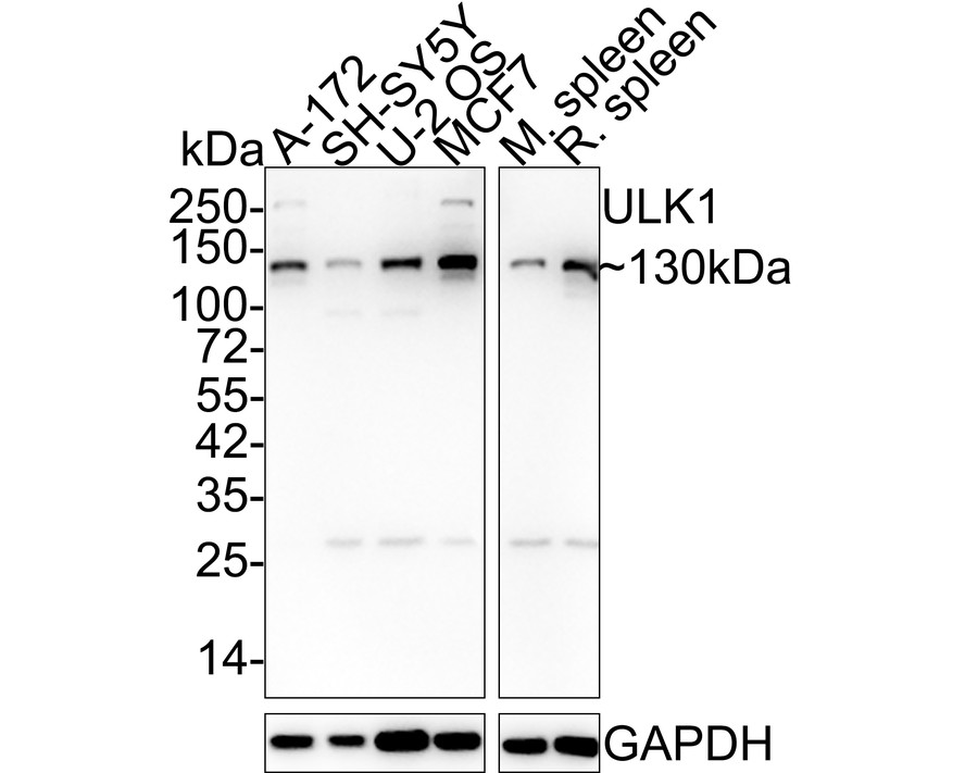
Western blot analysis of ULK1 on different lysates with Rabbit anti-ULK1 antibody at 1/5,000 dilution. Lane 1: A-172 cell lysate (15 µg/Lane), Lane 2: SH-SY5Y cell lysate (15 µg/Lane), Lane 3: U-2 OS cell lysate (15 µg/Lane), Lane 4: MCF7 cell lysate (15 µg/Lane), Lane 5: Mouse spleen tissue lysate (20 µg/Lane), Lane 6: Rat spleen tissue lysate (20 µg/Lane), Exposure time: 1 minute 30 seconds; 4-20% SDS-PAGE gel. Proteins were transferred to a PVDF membrane and blocked with 5% NFDM/TBST for 1 hour at room temperature. The primary antibody at 1/5,000 dilution was used in 5% NFDM/TBST at 4℃ overnight. Goat Anti-Rabbit IgG - HRP Secondary Antibody at 1/50,000 dilution was used for 1 hour at room temperature.WB
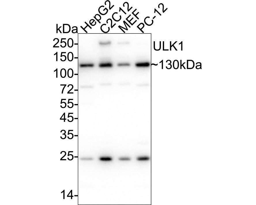
Western blot analysis of ULK1 on different lysates with Rabbit anti-ULK1 antibody at 1/5,000 dilution. Lane 1: HepG2 cell lysate, Lane 2: C2C12 cell lysate, Lane 3: MEF cell lysate, Lane 4: PC-12 cell lysate, Lysates/proteins at 20 µg/Lane. Exposure time: 6 seconds; 4-20% SDS-PAGE gel. Proteins were transferred to a PVDF membrane and blocked with 5% NFDM/TBST for 1 hour at room temperature. The primary antibody at 1/5,000 dilution was used in 5% NFDM/TBST at 4℃ overnight. Goat Anti-Rabbit IgG - HRP Secondary Antibody at 1/50,000 dilution was used for 1 hour at room temperature.IHC
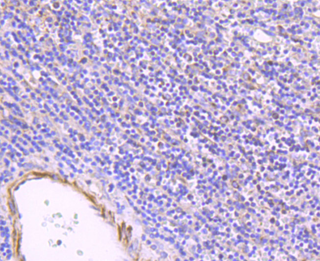
Immunohistochemical analysis of paraffin-embedded human spleen tissue using anti-ULK1 antibody. The section was pre-treated using heat mediated antigen retrieval with Tris-EDTA buffer (pH 8.0-8.4) for 20 minutes.The tissues were blocked in 5% BSA for 30 minutes at room temperature, washed with ddH2O and PBS, and then probed with the primary antibody (1/50) for 30 minutes at room temperature. The detection was performed using an HRP conjugated compact polymer system. DAB was used as the chromogen. Tissues were counterstained with hematoxylin and mounted with DPX.IHC
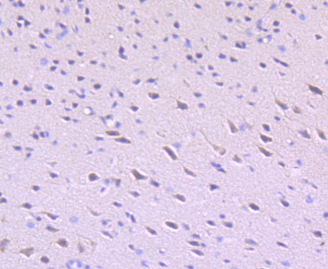
Immunohistochemical analysis of paraffin-embedded mouse brain tissue using anti-ULK1 antibody. The section was pre-treated using heat mediated antigen retrieval with Tris-EDTA buffer (pH 8.0-8.4) for 20 minutes.The tissues were blocked in 5% BSA for 30 minutes at room temperature, washed with ddH2O and PBS, and then probed with the primary antibody (1/50) for 30 minutes at room temperature. The detection was performed using an HRP conjugated compact polymer system. DAB was used as the chromogen. Tissues were counterstained with hematoxylin and mounted with DPX.IHC
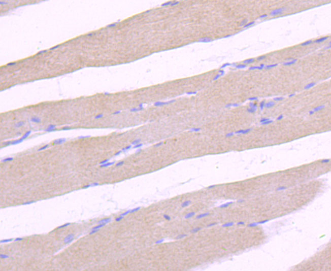
Immunohistochemical analysis of paraffin-embedded rat skeletal muscle tissue using anti-ULK1 antibody. The section was pre-treated using heat mediated antigen retrieval with Tris-EDTA buffer (pH 8.0-8.4) for 20 minutes.The tissues were blocked in 5% BSA for 30 minutes at room temperature, washed with ddH2O and PBS, and then probed with the primary antibody (1/50) for 30 minutes at room temperature. The detection was performed using an HRP conjugated compact polymer system. DAB was used as the chromogen. Tissues were counterstained with hematoxylin and mounted with DPX.| Product Name | ULK1 Recombinant Rabbit Monoclonal Antibody |
|---|---|
| Antibody Type | Primary Antibodies |
| Product description | Serine/threonine-protein kinase involved in autophagy in response to starvation. Acts upstream of phosphatidylinositol 3-kinase PIK3C3 to regulate the formation of autophagophores, the precursors of autophagosomes. Part of regulatory feedback loops in autophagy: acts both as a downstream effector and negative regulator of mammalian target of rapamycin complex 1 (mTORC1) via interaction with RPTOR. Activated via phosphorylation by AMPK and also acts as a regulator of AMPK by mediating phosphorylation of AMPK subunits PRKAA1, PRKAB2 and PRKAG1, leading to negatively regulate AMPK activity. May phosphorylate ATG13/KIAA0652 and RPTOR; however such data need additional evidences. Plays a role early in neuronal differentiation and is required for granule cell axon formation. May also phosphorylate SESN2 and SQSTM1 to regulate autophagy. |
| Immunogen | Synthetic peptide within Human ULK1 aa 930-979 / 1050. |
| Clonality | Monoclonal |
|---|---|
| Isotype | IgG |
| Host Species | Rabbit |
| Tested Applications | IHCWB |
| WB:1:5000 IHC:1:50-1:200 |
|
| Species Reactivity | HumanMouseRat |
| Concentration | 1mg/ml |
| Purification | Protein A |
| Alternative Names | ATG 1 antibody
ATG1 antibody
ATG1 autophagy related 1 homolog antibody
ATG1A antibody
Autophagy related protein 1 homolog antibody
Autophagy-related protein 1 homolog antibody
FLJ38455 antibody
FLJ46475 antibody
hATG1 antibody
KIAA0722 antibody
Serine/threonine protein kinase ULK1 antibody
Serine/threonine protein kinase Unc51.1 antibody
Serine/threonine-protein kinase ULK1 antibody
ULK 1 antibody
ULK1 antibody
ULK1_HUMAN antibody
Unc 51 (C. elegans) like kinase 1 antibody
UNC 51 antibody
Unc 51 like kinase 1 antibody
Unc-51 like kinase 1 (C. elegans) antibody
Unc-51-like kinase 1 antibody
UNC51 antibody
UNC51 C. elegans homolog of antibody Unc51.1 antibody |
|---|---|
| Molecular Weight(MW) | 113 kDa(Observed band size: 130 kDa) |
| Cellular Localization | Cytosol, Preautophagosomal structure. |
WB
Western blot analysis of ULK1 on different lysates with Rabbit anti-ULK1 antibody at 1/5,000 dilution. Lane 1: A-172 cell lysate (15 µg/Lane), Lane 2: SH-SY5Y cell lysate (15 µg/Lane), Lane 3: U-2 OS cell lysate (15 µg/Lane), Lane 4: MCF7 cell lysate (15 µg/Lane), Lane 5: Mouse spleen tissue lysate (20 µg/Lane), Lane 6: Rat spleen tissue lysate (20 µg/Lane), Exposure time: 1 minute 30 seconds; 4-20% SDS-PAGE gel. Proteins were transferred to a PVDF membrane and blocked with 5% NFDM/TBST for 1 hour at room temperature. The primary antibody at 1/5,000 dilution was used in 5% NFDM/TBST at 4℃ overnight. Goat Anti-Rabbit IgG - HRP Secondary Antibody at 1/50,000 dilution was used for 1 hour at room temperature.
WB
Western blot analysis of ULK1 on different lysates with Rabbit anti-ULK1 antibody at 1/5,000 dilution. Lane 1: HepG2 cell lysate, Lane 2: C2C12 cell lysate, Lane 3: MEF cell lysate, Lane 4: PC-12 cell lysate, Lysates/proteins at 20 µg/Lane. Exposure time: 6 seconds; 4-20% SDS-PAGE gel. Proteins were transferred to a PVDF membrane and blocked with 5% NFDM/TBST for 1 hour at room temperature. The primary antibody at 1/5,000 dilution was used in 5% NFDM/TBST at 4℃ overnight. Goat Anti-Rabbit IgG - HRP Secondary Antibody at 1/50,000 dilution was used for 1 hour at room temperature.
IHC
Immunohistochemical analysis of paraffin-embedded human spleen tissue using anti-ULK1 antibody. The section was pre-treated using heat mediated antigen retrieval with Tris-EDTA buffer (pH 8.0-8.4) for 20 minutes.The tissues were blocked in 5% BSA for 30 minutes at room temperature, washed with ddH2O and PBS, and then probed with the primary antibody (1/50) for 30 minutes at room temperature. The detection was performed using an HRP conjugated compact polymer system. DAB was used as the chromogen. Tissues were counterstained with hematoxylin and mounted with DPX.
IHC
Immunohistochemical analysis of paraffin-embedded mouse brain tissue using anti-ULK1 antibody. The section was pre-treated using heat mediated antigen retrieval with Tris-EDTA buffer (pH 8.0-8.4) for 20 minutes.The tissues were blocked in 5% BSA for 30 minutes at room temperature, washed with ddH2O and PBS, and then probed with the primary antibody (1/50) for 30 minutes at room temperature. The detection was performed using an HRP conjugated compact polymer system. DAB was used as the chromogen. Tissues were counterstained with hematoxylin and mounted with DPX.
IHC
Immunohistochemical analysis of paraffin-embedded rat skeletal muscle tissue using anti-ULK1 antibody. The section was pre-treated using heat mediated antigen retrieval with Tris-EDTA buffer (pH 8.0-8.4) for 20 minutes.The tissues were blocked in 5% BSA for 30 minutes at room temperature, washed with ddH2O and PBS, and then probed with the primary antibody (1/50) for 30 minutes at room temperature. The detection was performed using an HRP conjugated compact polymer system. DAB was used as the chromogen. Tissues were counterstained with hematoxylin and mounted with DPX.| Application Notes | WB:1:5000 IHC:1:50-1:200 |
|---|
| Form | Liquid |
|---|---|
| Storage Instructions | Store at +4℃ after thawing. Aliquot store at -20℃ or -80℃. Avoid repeated freeze / thaw cycles. |
| Storage Buffer | 1*TBS (pH7.4), 1%BSA, 40%Glycerol. Preservative: 0.05% Sodium Azide. |


