Application
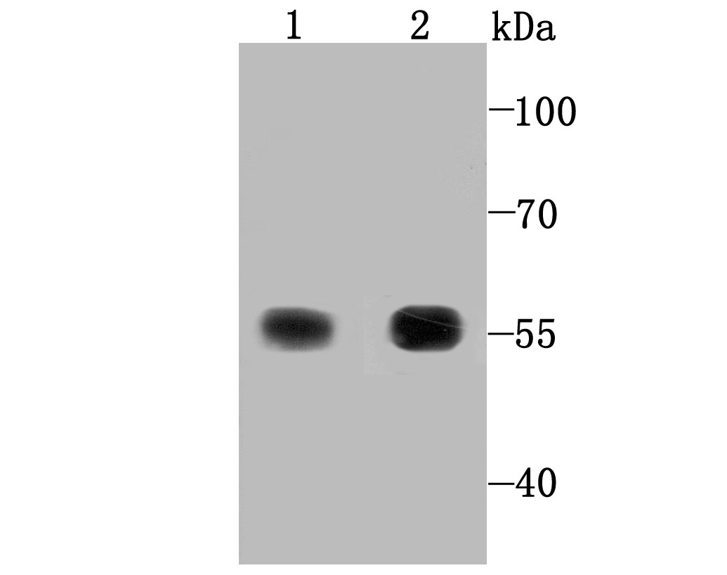
Fig1: Western blot analysis of TXNRD1 on Hela (1) and human liver tissue lysate (2) using anti-TXNRD1 antibody at 1/1,000 dilution.Application
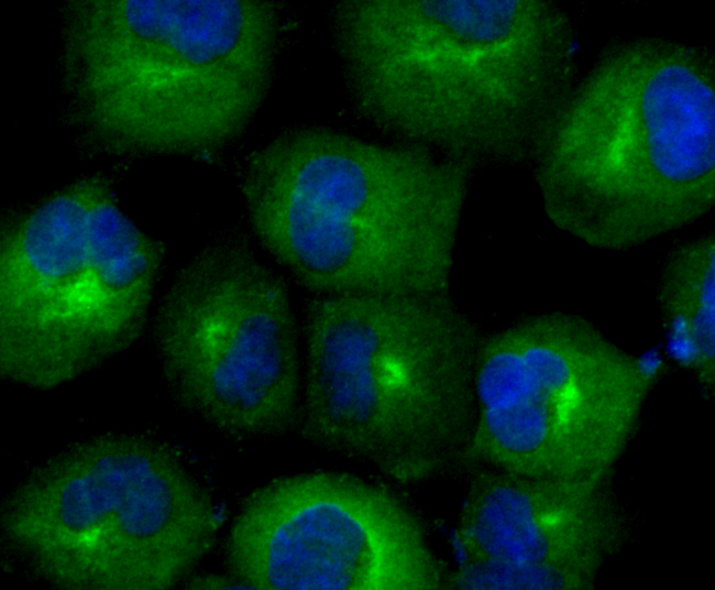
Fig2: ICC staining TXNRD1 in HUVEC cells (green). The nuclear counter stain is DAPI (blue). Cells were fixed in paraformaldehyde, permeabilised with 0.25% Triton X100/PBS.Application
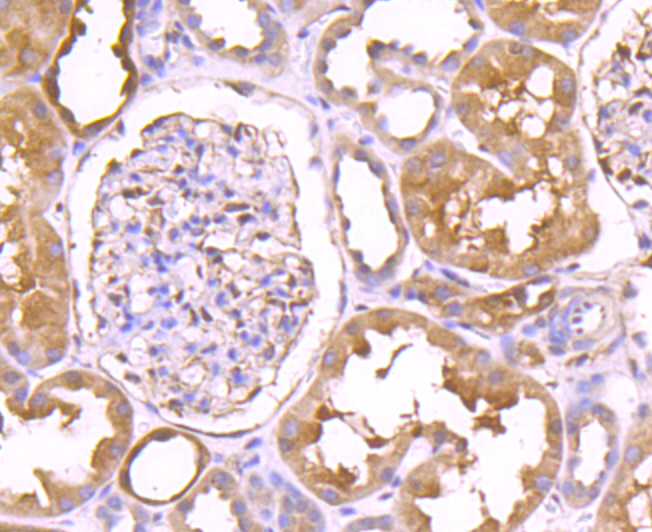
Fig3: Immunohistochemical analysis of paraffin-embedded human kidney tissue using anti-TXNRD1 antibody. Counter stained with hematoxylin.Application
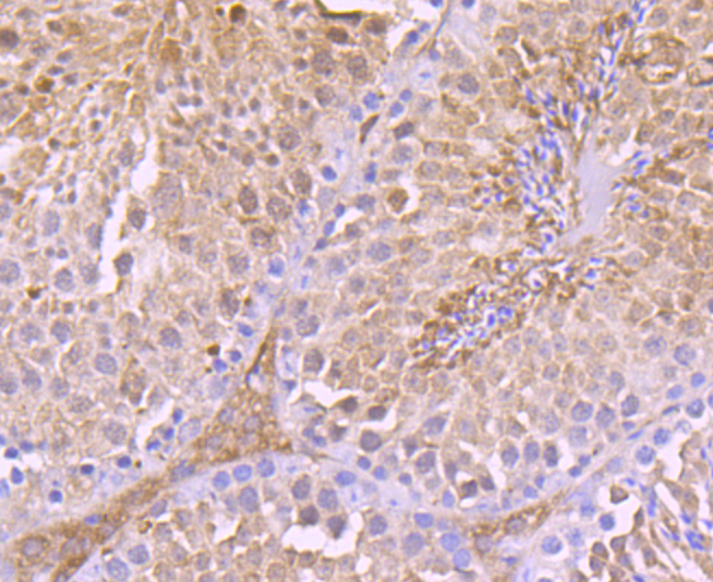
Fig4: Immunohistochemical analysis of paraffin-embedded mouse testes tissue using anti-TXNRD1 antibody. Counter stained with hematoxylin.Application
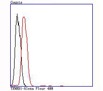
Fig5: Flow cytometric analysis of SH-SY5Y cells with TXNRD1 antibody at 1/100 dilution (red) compared with an unlabelled control (cells without incubation with primary antibody; black).| Product Name | TXNRD1 [JA11-32] |
|---|---|
| Antibody Type | Primary Antibodies |
| Antigen Alias | cytoplasmic antibody Gene associated with retinoic and IFN-induced mortality 12 protein antibody Gene associated with retinoic and interferon-induced mortality 12 protein antibody Gene associated with retinoid IFN induced mortality 12 protein antibody GRIM 12 antibody GRIM-12 antibody GRIM12 antibody KDRF antibody KM 102 derived reductase like factor antibody KM-102-derived reductase-like factor antibody MGC9145 antibody Oxidoreductase antibody Thioredoxin reductase 1 antibody Thioredoxin reductase 1 cytoplasmic antibody Thioredoxin reductase GRIM 12 antibody Thioredoxin reductase TR1 antibody TR 1 antibody TR antibody TR1 antibody TRXR 1 antibody TRXR1 antibody TRXR1_HUMAN antibody TXNR antibody TXNRD 1 antibody Txnrd1 antibody |
| Product description | Thioredoxin (Trx) is a redox protein that is found in several species, such as bacteria, plants and mammals, and contains a conserved active site, consisting of Trp-Cys-Gly-Pro-Cys. Trx reductases (TrxR1 and TrxR2) are ubiquitous Ly noble flavoproteins that catalyze the NADPH-dependent reduction of Trx as well as several other oxidized cellular components. Mammalian Trx reductases are a part of a selenium-containing pyridine nucleotide-disulphide oxidoreductase family, which has a conserved catalytic site of Cys-Val- Asn-Val-Gly-Cys. TrxR1 and TrxR2 are also involved in the prevention of oxidative stress. Inhibition of TrxR activity may provide for potential treatments of cancer, AIDS and other autoimmune diseases as well as bacterial infections and parasitic diseases. |
| Immunogen | recombinant protein |
| Clonality | Monoclonal |
|---|---|
| Isotype | IgG |
| Host Species | Recombinant rabbit |
| Tested Applications | WBICCIHCIPFC |
| WB:1:500-1:2,000 ICC:1:50-1:200 IHC:1:50-1:200 FC:1:50-1:100 |
|
| Species Reactivity | HumanMouseRat |
| Concentration | 1mg/ml |
| Alternative Names | cytoplasmic antibody Gene associated with retinoic and IFN-induced mortality 12 protein antibody Gene associated with retinoic and interferon-induced mortality 12 protein antibody Gene associated with retinoid IFN induced mortality 12 protein antibody GRIM 12 antibody GRIM-12 antibody GRIM12 antibody KDRF antibody KM 102 derived reductase like factor antibody KM-102-derived reductase-like factor antibody MGC9145 antibody Oxidoreductase antibody Thioredoxin reductase 1 antibody Thioredoxin reductase 1 cytoplasmic antibody Thioredoxin reductase GRIM 12 antibody Thioredoxin reductase TR1 antibody TR 1 antibody TR antibody TR1 antibody TRXR 1 antibody TRXR1 antibody TRXR1_HUMAN antibody TXNR antibody TXNRD 1 antibody Txnrd1 antibody |
|---|---|
| Molecular Weight(MW) | 55 kDa |
| Cellular Localization | Cytoplasm. Nucleus. |
Application
Fig1: Western blot analysis of TXNRD1 on Hela (1) and human liver tissue lysate (2) using anti-TXNRD1 antibody at 1/1,000 dilution.
Application
Fig2: ICC staining TXNRD1 in HUVEC cells (green). The nuclear counter stain is DAPI (blue). Cells were fixed in paraformaldehyde, permeabilised with 0.25% Triton X100/PBS.
Application
Fig3: Immunohistochemical analysis of paraffin-embedded human kidney tissue using anti-TXNRD1 antibody. Counter stained with hematoxylin.
Application
Fig4: Immunohistochemical analysis of paraffin-embedded mouse testes tissue using anti-TXNRD1 antibody. Counter stained with hematoxylin.
Application
Fig5: Flow cytometric analysis of SH-SY5Y cells with TXNRD1 antibody at 1/100 dilution (red) compared with an unlabelled control (cells without incubation with primary antibody; black).| Positive Control | Human liver tissue lysate, Hela, HUVEC, SH-SY5Y, mouse testes tissue, human kidney tissue. |
|---|---|
| Application Notes | WB:1:500-1:2,000 ICC:1:50-1:200 IHC:1:50-1:200 FC:1:50-1:100 |
| Form | Liquid |
|---|---|
| Storage Instructions | Store at +4℃ after thawing. Aliquot store at -20℃ or -80℃. Avoid repeated freeze / thaw cycles. |
| Storage Buffer | 1*TBS (pH7.4), 1%BSA, 40%Glycerol. Preservative: 0.05% Sodium Azide. |


