WB
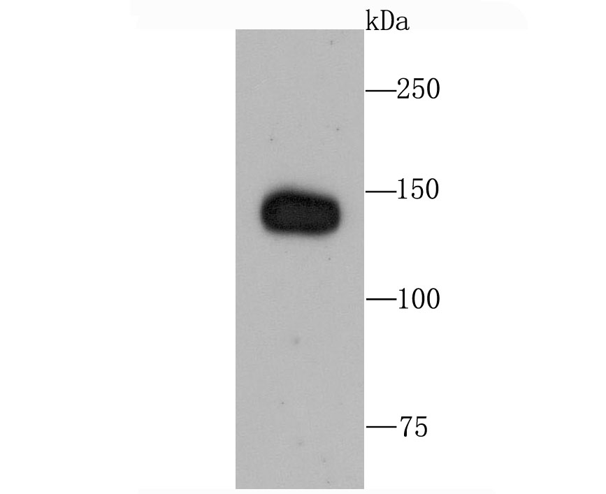
Western blot analysis of Factor H on human lung tissue lysate using anti-Factor H antibody at 1/1,000 dilution.ICC/IF
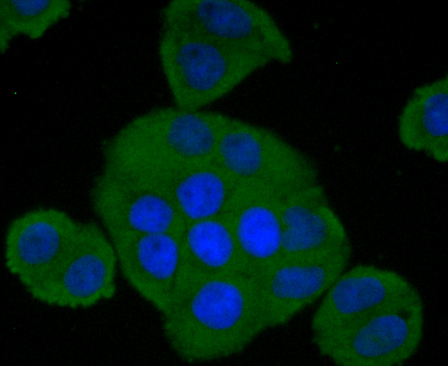
ICC staining Factor H in Hela cells (green). The nuclear counter stain is DAPI (blue). Cells were fixed in paraformaldehyde, permeabilised with 0.25% Triton X100/PBS.ICC/IF
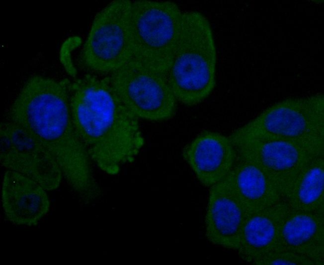
ICC staining Factor H in HepG2 cells (green). The nuclear counter stain is DAPI (blue). Cells were fixed in paraformaldehyde, permeabilised with 0.25% Triton X100/PBS.IHC
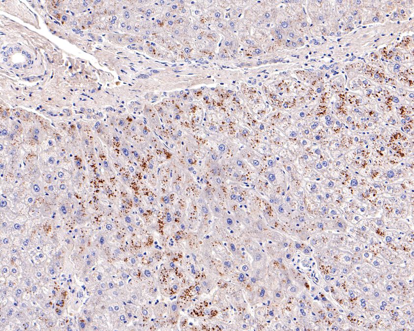
Immunohistochemical analysis of paraffin-embedded human liver tissue using anti-Factor H antibody. The section was pre-treated using heat mediated antigen retrieval with Tris-EDTA buffer (pH 8.0-8.4) for 20 minutes.The tissues were blocked in 5% BSA for 30 minutes at room temperature, washed with ddH2O and PBS, and then probed with the primary antibody (1/100) for 30 minutes at room temperature. The detection was performed using an HRP conjugated compact polymer system. DAB was used as the chromogen. Tissues were counterstained with hematoxylin and mounted with DPX.| Product Name | Factor H Recombinant Rabbit Monoclonal Antibody |
|---|---|
| Antibody Type | Primary Antibodies |
| Product description | The Factor H gene family is a multidomain, multifunctional protein family whose individual members are defined by conserved structural elements, which display diverse yet often overlapping functions. These proteins share a common structural motif, the short consensus repeat (SCR), which is structurally conserved among related genes and between phylogenetically divergent species. The human complement Factor H (FH, CFH, HUS, b-1H) gene encodes a 1213 amino acid serum glycoprotein which is arranged into 20 SCRs, each approximately 60 amino acids long, and an 18-residue leader sequence. Factor H controls the function of the alternative complement pathway and acts as a cofactor with Factor I (C3b inactivator). In addition, Factor H has functional activity outside of the complement system, where it can bind to the cellular integrin receptor (CD11b/CD18), interact with cell surface glycosaminoglycans and associate with the surface of certain pathogenic microorganisms. Deficiencies in Factor H is a common characteristic of acute renal disease. |
| Immunogen | Synthetic peptide within Human Factor H aa 131-180 / 1231. |
| Clonality | Monoclonal |
|---|---|
| Isotype | IgG |
| Host Species | Recombinant rabbit |
| Tested Applications | ICC/IFIHCWB |
| WB:1:500-1:2000 IHC:1:50-1:100 ICC/IF:1:50-1:100 |
|
| Species Reactivity | Human |
| Concentration | 1mg/ml |
| Purification | Protein A |
| Alternative Names | adrenomedullin binding protein antibody
age related maculopathy susceptibility 1 antibody
AHUS 1 antibody
AHUS1 antibody
AMBP 1 antibody
AMBP1 antibody
ARMD 4 antibody
ARMD4 antibody
ARMS 1 antibody
ARMS1 antibody
beta 1 H globulin antibody
beta 1H antibody
beta1H antibody
CFAH_HUMAN antibody
CFH antibody
CFHL 3 antibody
CFHL3 antibody
Complement factor H antibody
complement factor H isoform b antibody Factor H antibody factor H like 1 antibody FH antibody FHL 1 antibody FHL1 antibody H factor 1 (complement) antibody H factor 1 antibody H factor 2 (complement) antibody HF 1 antibody HF 2 antibody HF antibody HF1 antibody HF2 antibody HUS antibody MGC88246 antibody |
|---|---|
| Molecular Weight(MW) | 140 kDa |
| Cellular Localization | Secreted. |
| SwissProt ID | P08603 |
|---|
WB
Western blot analysis of Factor H on human lung tissue lysate using anti-Factor H antibody at 1/1,000 dilution.
ICC/IF
ICC staining Factor H in Hela cells (green). The nuclear counter stain is DAPI (blue). Cells were fixed in paraformaldehyde, permeabilised with 0.25% Triton X100/PBS.
ICC/IF
ICC staining Factor H in HepG2 cells (green). The nuclear counter stain is DAPI (blue). Cells were fixed in paraformaldehyde, permeabilised with 0.25% Triton X100/PBS.
IHC
Immunohistochemical analysis of paraffin-embedded human liver tissue using anti-Factor H antibody. The section was pre-treated using heat mediated antigen retrieval with Tris-EDTA buffer (pH 8.0-8.4) for 20 minutes.The tissues were blocked in 5% BSA for 30 minutes at room temperature, washed with ddH2O and PBS, and then probed with the primary antibody (1/100) for 30 minutes at room temperature. The detection was performed using an HRP conjugated compact polymer system. DAB was used as the chromogen. Tissues were counterstained with hematoxylin and mounted with DPX.| Application Notes | WB:1:500-1:2000 IHC:1:50-1:100 ICC/IF:1:50-1:100 |
|---|
| Form | Liquid |
|---|---|
| Storage Instructions | Store at +4℃ after thawing. Aliquot store at -20℃ or -80℃. Avoid repeated freeze / thaw cycles. |
| Storage Buffer | 1*TBS (pH7.4), 1%BSA, 40%Glycerol. Preservative: 0.05% Sodium Azide. |


