IHC
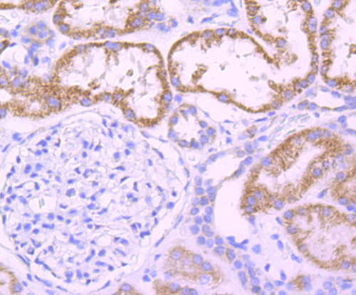
Immunohistochemical analysis of paraffin-embedded human kidney tissue using anti-DOPA Decarboxylase antibody. Counter stained with hematoxylin.IHC
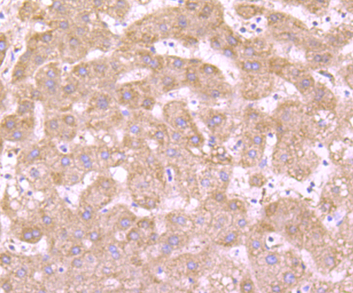
Immunohistochemical analysis of paraffin-embedded human liver tissue using anti-DOPA Decarboxylase antibody. Counter stained with hematoxylin.WB
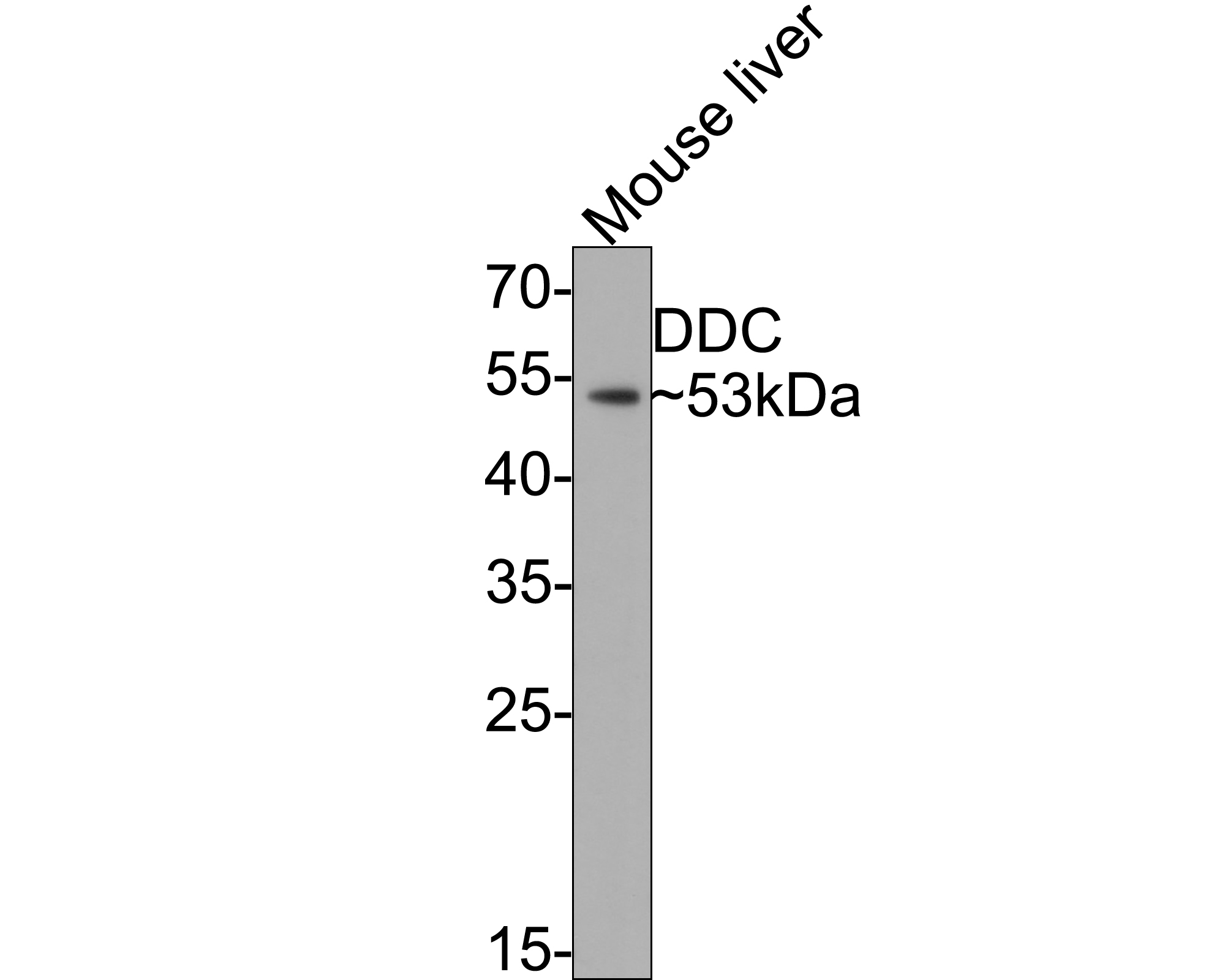
Western blot analysis of DOPA Decarboxylase on mouse liver tissue lysates with Rabbit anti-DOPA Decarboxylase antibody at 1/500 dilution. Lysates/proteins at 20 µg/Lane. Exposure time: 2 minutes; 10% SDS-PAGE gel. Proteins were transferred to a PVDF membrane and blocked with 5% NFDM/TBST for 1 hour at room temperature. The primary antibody at 1/500 dilution was used in 5% NFDM/TBST at room temperature for 2 hours. Goat Anti-Rabbit IgG - HRP Secondary Antibody at 1:200,000 dilution was used for 1 hour at room temperature.WB
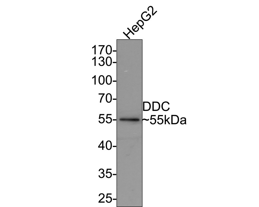
Western blot analysis of DOPA Decarboxylase on HepG2 cell lysates with Rabbit anti-DOPA Decarboxylase antibody at 1/500 dilution. Lysates/proteins at 10 µg/Lane. Exposure time: 2 minutes; 10% SDS-PAGE gel. Proteins were transferred to a PVDF membrane and blocked with 5% NFDM/TBST for 1 hour at room temperature. The primary antibody at 1/500 dilution was used in 5% NFDM/TBST at room temperature for 2 hours. Goat Anti-Rabbit IgG - HRP Secondary Antibody at 1:200,000 dilution was used for 1 hour at room temperature.| Product Name | DOPA Decarboxylase Recombinant Rabbit Monoclonal Antibody |
|---|---|
| Antibody Type | Primary Antibodies |
| Product description | DOPA decarboxylase (DDC), also designated aromatic-L-amino-acid decarboxylase (AADC) belongs to the group II decarboxylase family of proteins. DDC, which can form a homodimer, is an important protein in the catecholamine biosynthesis pathway. DDC acts as a catalyst in the decarboxylation of L-5-hydroxytryptophan to serotonin, L-3,4-dihydroxyphenylalanine (DOPA) to dopamine and L-tryptophan to tryptamine. Defects in the gene encoding for DDC may cause the autosomal recessive disorder AADC deficiency. AADC deficiency is an early onset inborn error in neurotransmitter metabolism which can lead to catecholamine and serotonin deficiency. This causes poor feeding, psychomotor and developmental delays, lethargy, ptosis, gastrointestinal disturbances and hypothermia. |
| Immunogen | Synthetic peptide within Human DOPA Decarboxylase aa 27-76 / 480. |
| Clonality | monoclonal |
|---|---|
| Isotype | IgG |
| Host Species | Recombinant rabbit |
| Tested Applications | IHCWB |
| WB:1:500-1:1000 IHC:1:50-1:400 |
|
| Species Reactivity | HumanMouse |
| Concentration | 1mg/ml |
| Purification | Protein A |
| Alternative Names | AADC antibody Aromatic L Amino Acid Decarboxylase antibody Aromatic-L-amino-acid decarboxylase antibody Ddc antibody DDC_HUMAN antibody DOPA decarboxylase (aromatic L-amino acid decarboxylase) antibody DOPA decarboxylase antibody |
|---|---|
| Molecular Weight(MW) | 53 kDa |
| Cellular Localization | Cytoplasm. |
IHC
Immunohistochemical analysis of paraffin-embedded human kidney tissue using anti-DOPA Decarboxylase antibody. Counter stained with hematoxylin.
IHC
Immunohistochemical analysis of paraffin-embedded human liver tissue using anti-DOPA Decarboxylase antibody. Counter stained with hematoxylin.
WB
Western blot analysis of DOPA Decarboxylase on mouse liver tissue lysates with Rabbit anti-DOPA Decarboxylase antibody at 1/500 dilution. Lysates/proteins at 20 µg/Lane. Exposure time: 2 minutes; 10% SDS-PAGE gel. Proteins were transferred to a PVDF membrane and blocked with 5% NFDM/TBST for 1 hour at room temperature. The primary antibody at 1/500 dilution was used in 5% NFDM/TBST at room temperature for 2 hours. Goat Anti-Rabbit IgG - HRP Secondary Antibody at 1:200,000 dilution was used for 1 hour at room temperature.
WB
Western blot analysis of DOPA Decarboxylase on HepG2 cell lysates with Rabbit anti-DOPA Decarboxylase antibody at 1/500 dilution. Lysates/proteins at 10 µg/Lane. Exposure time: 2 minutes; 10% SDS-PAGE gel. Proteins were transferred to a PVDF membrane and blocked with 5% NFDM/TBST for 1 hour at room temperature. The primary antibody at 1/500 dilution was used in 5% NFDM/TBST at room temperature for 2 hours. Goat Anti-Rabbit IgG - HRP Secondary Antibody at 1:200,000 dilution was used for 1 hour at room temperature.| Application Notes | WB:1:500-1:1000 IHC:1:50-1:400 |
|---|
| Form | Liquid |
|---|---|
| Storage Instructions | Store at +4℃ after thawing. Aliquot store at -20℃ or -80℃. Avoid repeated freeze / thaw cycles. |
| Storage Buffer | 1*TBS (pH7.4), 1%BSA, 40%Glycerol. Preservative: 0.05% Sodium Azide. |


