Application
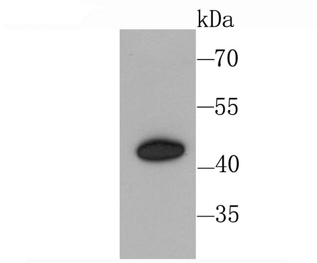
Fig1: Western blot analysis of CXCR3 on Hela cell using anti-CXCR3 antibody at 1/1,000 dilution.Application
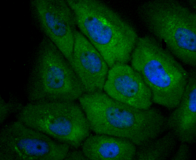
Fig2: ICC staining CXCR3 in A431 cells (green). The nuclear counter stain is DAPI (blue). Cells were fixed in paraformaldehyde, permeabilised with 0.25% Triton X100/PBS.Application
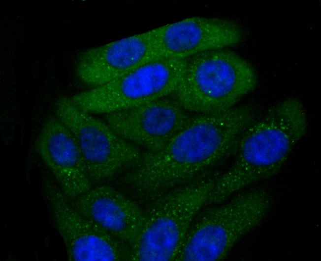
Fig3: ICC staining CXCR3 in HepG2 cells (green). The nuclear counter stain is DAPI (blue). Cells were fixed in paraformaldehyde, permeabilised with 0.25% Triton X100/PBS.Application
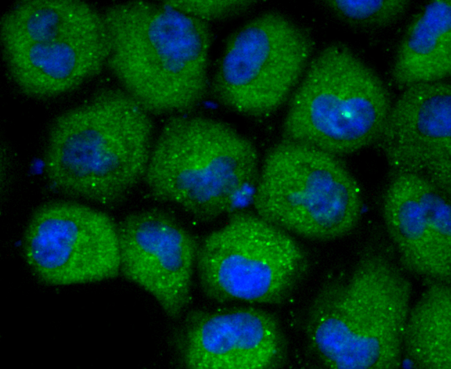
Fig4: ICC staining CXCR3 in HUVEC cells (green). The nuclear counter stain is DAPI (blue). Cells were fixed in paraformaldehyde, permeabilised with 0.25% Triton X100/PBS.| Product Name | CXCR3 [JA61-33] |
|---|---|
| Antibody Type | Primary Antibodies |
| Antigen Alias | C-X-C chemokine receptor type 3 antibody CD 183 antibody CD182 antibody CD183 antibody Chemokine (C X C motif) receptor 3 antibody Chemokine (C X C) receptor 3 antibody Chemokine CXC Motif Receptor 3 antibody CKR L2 antibody CKR-L2 antibody CKRL2 antibody CMKAR3 antibody CXC-R3 antibody CXCR-3 antibody CXCR3 antibody CXCR3_HUMAN antibody G Protein Coupled Receptor 9 antibody G protein-coupled receptor 9 antibody GPR9 antibody Interferon-inducible protein 10 receptor antibody IP-10 receptor antibody IP10 antibody IP10 R antibody IP10 receptor antibody IP10-R antibody IP10R antibody Mig R antibody Mig receptor antibody Mig-R antibody MIGR antibody |
| Product description | The C-X-C or α chemokine family is characterized by a pair of cysteine residues separated by a single amino acid and primarily functions as chemoattractants for neutrophils. The C-X-C family includes IL-8, NAP-2, MSGA and stromal cell derived factor-1 (SDF-1). SDF-1 was originally described as a pre-B cell stimulatory factor, but has since been shown to function as a potent chemoattractant for T cells and monocytes but not neutrophils. Receptors for the C-X-C family are G protein-coupled, seven pass transmembrane domain proteins which include IL-8RA, IL-8RB, CXCR-3 and fusin (also designated LESTR or CXCR-4). CXCR-3, also known as IP-10/MIG receptor, mediates Ca2+ mobilization and chemotaxis in response to the C-X-C chemokines IP-10 and MIG. CXCR-3 is highly expressed in IL-2-activated T lymphocytes, but not in resting T lymphocytes, B lymphocytes, monocytes or granulocytes. |
| Immunogen | recombinant protein |
| Clonality | Monoclonal |
|---|---|
| Isotype | IgG |
| Host Species | Recombinant rabbit |
| Tested Applications | WBICC/IFIP |
| WB:1:500-1:2,000 ICC:1:50-1:200 |
|
| Species Reactivity | Human |
| Concentration | 1mg/ml |
| Alternative Names | C-X-C chemokine receptor type 3 antibody CD 183 antibody CD182 antibody CD183 antibody Chemokine (C X C motif) receptor 3 antibody Chemokine (C X C) receptor 3 antibody Chemokine CXC Motif Receptor 3 antibody CKR L2 antibody CKR-L2 antibody CKRL2 antibody CMKAR3 antibody CXC-R3 antibody CXCR-3 antibody CXCR3 antibody CXCR3_HUMAN antibody G Protein Coupled Receptor 9 antibody G protein-coupled receptor 9 antibody GPR9 antibody Interferon-inducible protein 10 receptor antibody IP-10 receptor antibody IP10 antibody IP10 R antibody IP10 receptor antibody IP10-R antibody IP10R antibody Mig R antibody Mig receptor antibody Mig-R antibody MIGR antibody |
|---|---|
| Molecular Weight(MW) | 41 kDa |
| Cellular Localization | Cell membrane. |
| SwissProt ID | P49682 |
|---|
Application
Fig1: Western blot analysis of CXCR3 on Hela cell using anti-CXCR3 antibody at 1/1,000 dilution.
Application
Fig2: ICC staining CXCR3 in A431 cells (green). The nuclear counter stain is DAPI (blue). Cells were fixed in paraformaldehyde, permeabilised with 0.25% Triton X100/PBS.
Application
Fig3: ICC staining CXCR3 in HepG2 cells (green). The nuclear counter stain is DAPI (blue). Cells were fixed in paraformaldehyde, permeabilised with 0.25% Triton X100/PBS.
Application
Fig4: ICC staining CXCR3 in HUVEC cells (green). The nuclear counter stain is DAPI (blue). Cells were fixed in paraformaldehyde, permeabilised with 0.25% Triton X100/PBS.| Positive Control | Hela, A431, HepG2, HUVEC. |
|---|---|
| Application Notes | WB:1:500-1:2,000 ICC:1:50-1:200 |
| Form | Liquid |
|---|---|
| Storage Instructions | Store at +4℃ after thawing. Aliquot store at -20℃ or -80℃. Avoid repeated freeze / thaw cycles. |
| Storage Buffer | 1*TBS (pH7.4), 1%BSA, 40%Glycerol. Preservative: 0.05% Sodium Azide. |


