Application
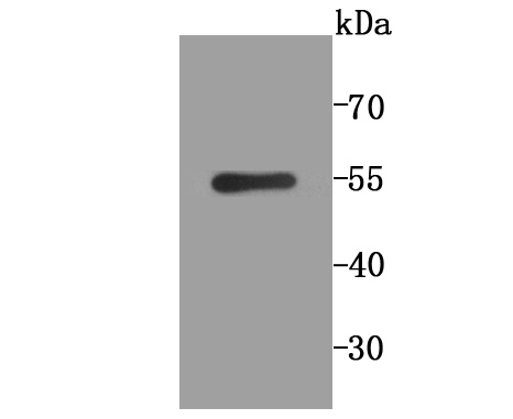
Fig1: Western blot analysis of PLGF on SH-SY5Y cell lysate using anti-PLGF antibody at 1/1,000 dilution.Application
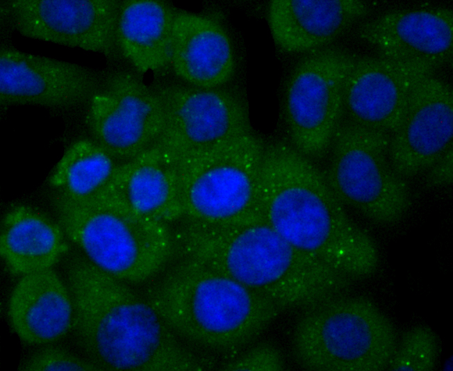
Fig2: ICC staining PLGF in HepG2 cells (green). The nuclear counter stain is DAPI (blue). Cells were fixed in paraformaldehyde, permeabilised with 0.25% Triton X100/PBS.Application
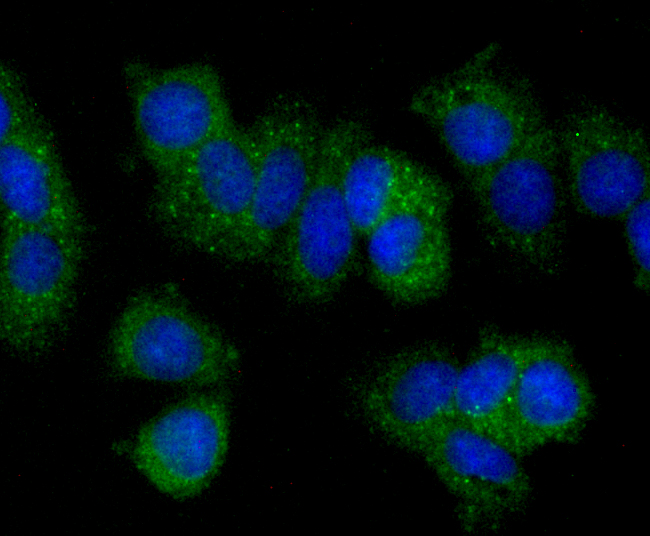
Fig3: ICC staining PLGF in MCF-7 cells (green). The nuclear counter stain is DAPI (blue). Cells were fixed in paraformaldehyde, permeabilised with 0.25% Triton X100/PBS.Application
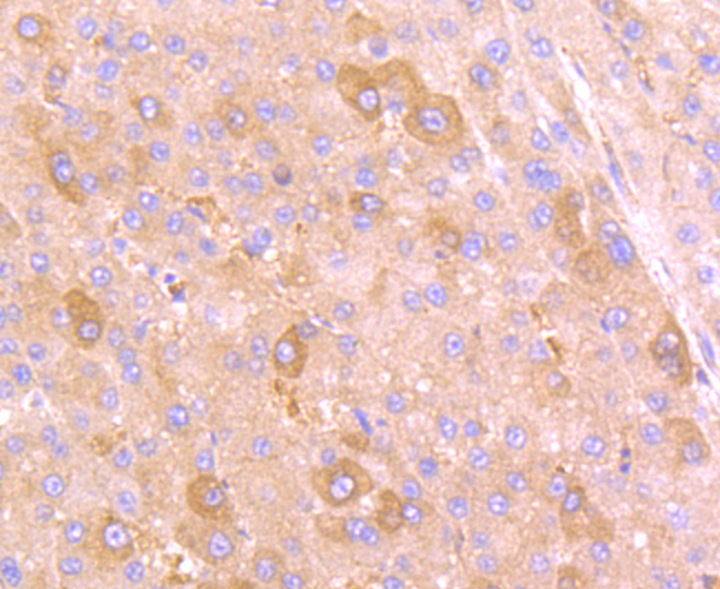
Fig4: Immunohistochemical analysis of paraffin-embedded human liver cancer tissue using anti-PLGF antibody. Counter stained with hematoxylin.Application
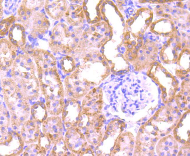
Fig5: Immunohistochemical analysis of paraffin-embedded rat kidney tissue using anti-PLGF antibody. Counter stained with hematoxylin.Application
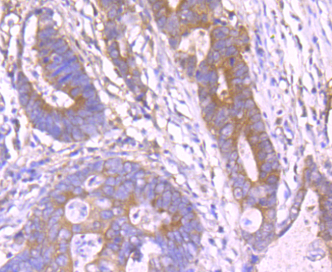
Fig6: Immunohistochemical analysis of paraffin-embedded human colon cancer tissue using anti-PLGF antibody. Counter stained with hematoxylin.Application
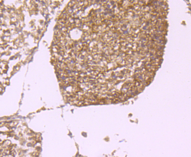
Fig7: Immunohistochemical analysis of paraffin-embedded mouse orchis tissue using anti-PLGF antibody. Counter stained with hematoxylin.| Product Name | PLGF [JA63-15] |
|---|---|
| Antibody Type | Primary Antibodies |
| Antigen Alias | D12S1900 antibody Pgf antibody PGFL antibody PIGF antibody Placenta growth factor antibody Placental growth factor antibody Placental growth factor, vascular endothelial growth factor related protein antibody PlGF 2 antibody PlGF antibody PLGF_HUMAN antibody PlGF2 antibody SHGC 10760 antibody |
| Product description | he onset of angiogenesis is believed to be an early event in tumorigenesis and may facilitate tumor progression and metastasis. Several growth factors with angiogenic activity have been described. These include fibroblast growth factor (FGF), platelet derived growth factor (PDGF), vascular endothelial growth factor (VEGF) and placenta growth factor (PlGF). Like VEGF, several PlGF variants have been shown to arise from alternative mRNA splicings. Evidence has suggested VEGF to be an obligatory component in PlGF signaling. While VEGF homodimers and VEGF/PlGF heterodimers function as potent mediators of mitogenic and chemotactic responses in endothelial cells, PlGF homodimers are effectual only at extremely high concentrations. Indeed, many of the physiological effects attributed to VEGF may actually be a result of VEGF/PlGF. VEGF and PlGF share a common receptor, Flt-1, and may also activate Flk-1/KDR. |
| Immunogen | Recombinant protein |
| Clonality | Monoclonal |
|---|---|
| Isotype | IgG |
| Host Species | Recombinant rabbit |
| Tested Applications | WBICCIHC |
| WB:1:500~1:2000 ICC:1:200~1:500 IHC:1:50~1:200 Notes:Optimal dilutions/concentrations should be determined by the researcher. |
|
| Species Reactivity | Human |
| Concentration | 1mg/ml |
| Alternative Names | D12S1900 antibody
Pgf antibody
PGFL antibody
PIGF antibody
Placenta growth factor antibody
Placental growth factor antibody
Placental growth factor vascular endothelial growth factor related protein antibody PlGF 2 antibody PlGF antibody PLGF_HUMAN antibody PlGF2 antibody SHGC 10760 antibody |
|---|---|
| Molecular Weight(MW) | 50 kDa |
| Cellular Localization | Secreted. |
| SwissProt ID | P49763 |
|---|
Application
Fig1: Western blot analysis of PLGF on SH-SY5Y cell lysate using anti-PLGF antibody at 1/1,000 dilution.
Application
Fig2: ICC staining PLGF in HepG2 cells (green). The nuclear counter stain is DAPI (blue). Cells were fixed in paraformaldehyde, permeabilised with 0.25% Triton X100/PBS.
Application
Fig3: ICC staining PLGF in MCF-7 cells (green). The nuclear counter stain is DAPI (blue). Cells were fixed in paraformaldehyde, permeabilised with 0.25% Triton X100/PBS.
Application
Fig4: Immunohistochemical analysis of paraffin-embedded human liver cancer tissue using anti-PLGF antibody. Counter stained with hematoxylin.
Application
Fig5: Immunohistochemical analysis of paraffin-embedded rat kidney tissue using anti-PLGF antibody. Counter stained with hematoxylin.
Application
Fig6: Immunohistochemical analysis of paraffin-embedded human colon cancer tissue using anti-PLGF antibody. Counter stained with hematoxylin.
Application
Fig7: Immunohistochemical analysis of paraffin-embedded mouse orchis tissue using anti-PLGF antibody. Counter stained with hematoxylin.| Positive Control | SH-SY5Y cell lysate, HepG2, MCF-7, rat kidney tissue, human colon cancer tissue, mouse orchis tissue, human liver cancer tissue. |
|---|---|
| Application Notes | WB:1:500~1:2000 ICC:1:200~1:500 IHC:1:50~1:200 Notes:Optimal dilutions/concentrations should be determined by the researcher. |
| Form | Liquid |
|---|---|
| Storage Instructions | Store at +4℃ after thawing. Aliquot store at -20℃ or -80℃. Avoid repeated freeze / thaw cycles. |
| Storage Buffer | 1*TBS (pH7.4), 1%BSA, 40%Glycerol. Preservative: 0.05% Sodium Azide. |


