WB
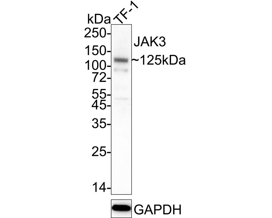
Western blot analysis of JAK3 on TF-1 cell lysates with Rabbit anti-JAK3 antibody at 1/1,000 dilution. Lysates/proteins at 20 µg/Lane. Exposure time: 3 minutes; 4-20% SDS-PAGE gel. Proteins were transferred to a PVDF membrane and blocked with 5% NFDM/TBST for 1 hour at room temperature. The primary antibody at 1/1,000 dilution was used in 5% NFDM/TBST at 4℃ overnight. Goat Anti-Rabbit IgG - HRP Secondary Antibody at 1/50,000 dilution was used for 1 hour at room temperature.IHC
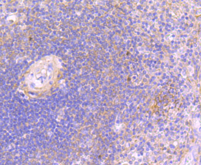
Immunohistochemical analysis of paraffin-embedded human spleen tissue using anti-JAK3 antibody. Counter stained with hematoxylin.IHC
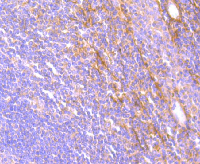
Immunohistochemical analysis of paraffin-embedded human tonsil tissue using anti-JAK3 antibody. Counter stained with hematoxylin.ICC/IF
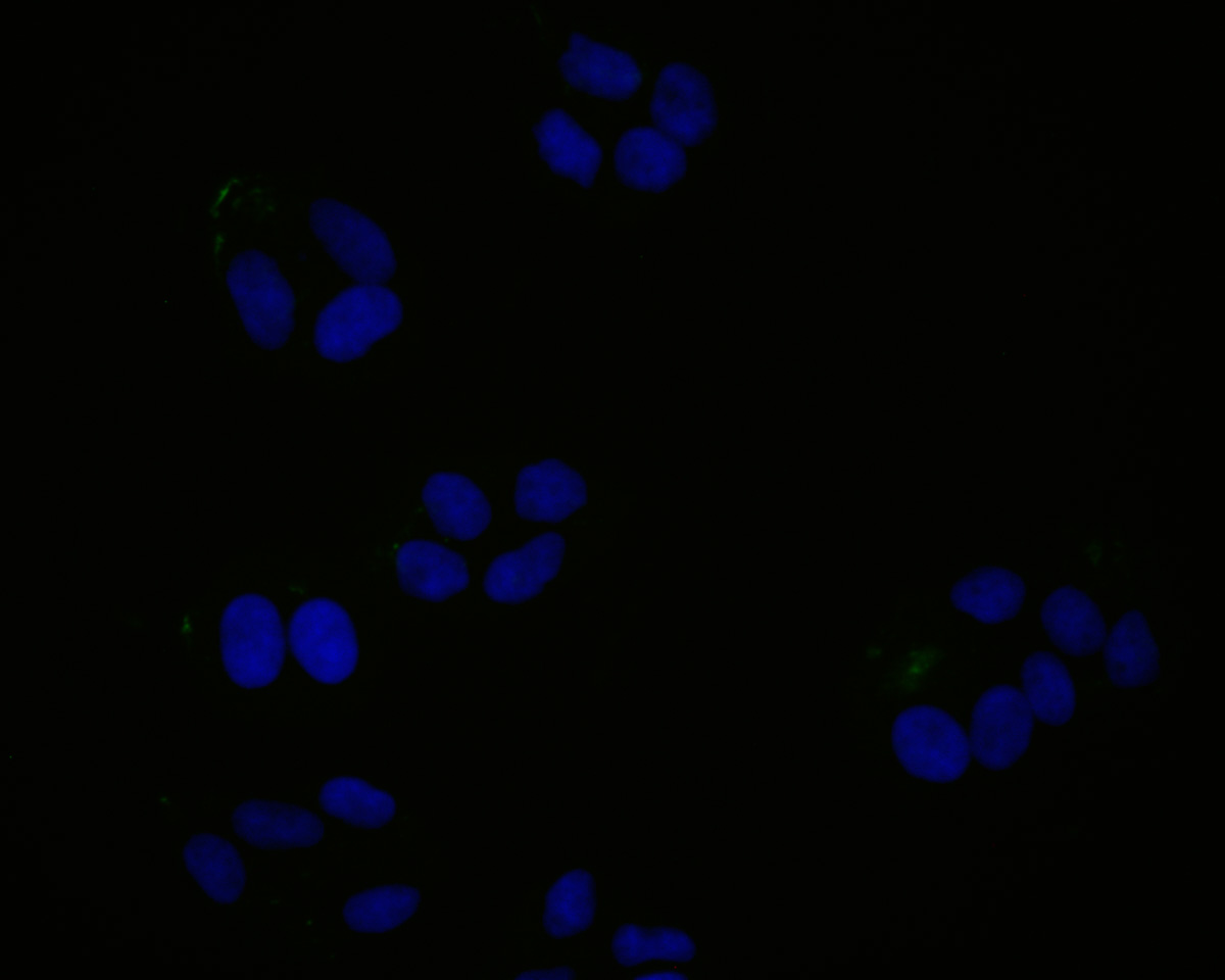
ICC staining of JAK3 in Hela cells (green). Formalin fixed cells were permeabilized with 0.1% Triton X-100 in TBS for 10 minutes at room temperature and blocked with 1% Blocker BSA for 15 minutes at room temperature. Cells were probed with the primary antibody (1/50) for 1 hour at room temperature, washed with PBS. Alexa Fluor®488 Goat anti-Rabbit IgG was used as the secondary antibody at 1/1,000 dilution. The nuclear counter stain is DAPI (blue).| Product Name | JAK3 Recombinant Rabbit Monoclonal Antibody |
|---|---|
| Antibody Type | Primary Antibodies |
| Product description | Non-receptor tyrosine kinase involved in various processes such as cell growth, development, or differentiation. Mediates essential signaling events in both innate and adaptive immunity and plays a crucial role in hematopoiesis during T-cells development. In the cytoplasm, plays a pivotal role in signal transduction via its association with type I receptors sharing the common subunit gamma such as IL2R, IL4R, IL7R, IL9R, IL15R and IL21R. Following ligand binding to cell surface receptors, phosphorylates specific tyrosine residues on the cytoplasmic tails of the receptor, creating docking sites for STATs proteins. Subsequently, phosphorylates the STATs proteins once they are recruited to the receptor. Phosphorylated STATs then form homodimer or heterodimers and translocate to the nucleus to activate gene transcription. For example, upon IL2R activation by IL2, JAK1 and JAK3 molecules bind to IL2R beta (IL2RB) and gamma chain (IL2RG) subunits inducing the tyrosine phosphorylation of both receptor subunits on their cytoplasmic domain. Then, STAT5A AND STAT5B are recruited, phosphorylated and activated by JAK1 and JAK3. |
| Immunogen | Synthetic peptide within N-terminal human JAK3. |
| Clonality | monoclonal |
|---|---|
| Isotype | IgG |
| Host Species | Recombinant rabbit |
| Tested Applications | ICC/IFIHCWB |
| WB:1:1000 IHC:1:50-1:200 ICC/IF:1:50-1:100 |
|
| Species Reactivity | Human |
| Concentration | 1mg/ml |
| Purification | Protein A |
| Alternative Names | EC 2.7.10.2 antibody
JAK 3 antibody
JAK L antibody JAK-3 antibody Jak3 antibody JAK3 HUMAN antibody JAK3_HUMAN antibody JAKL antibody Janus kinase 3 (a protein tyrosine kinase leukocyte) antibody Janus kinase 3 antibody Janus Kinase3 antibody L JAK antibody L-JAK antibody Leukocyte janus kinase antibody LJAK antibody Protein tyrosine kinase leukocyte antibody Tyrosine protein kinase JAK3 antibody Tyrosine-protein kinase JAK3 antibody |
|---|---|
| Molecular Weight(MW) | 125kDa |
| Cellular Localization | Endomembrane system, Cytoplasm. |
| SwissProt ID | P52333 |
|---|
WB
Western blot analysis of JAK3 on TF-1 cell lysates with Rabbit anti-JAK3 antibody at 1/1,000 dilution. Lysates/proteins at 20 µg/Lane. Exposure time: 3 minutes; 4-20% SDS-PAGE gel. Proteins were transferred to a PVDF membrane and blocked with 5% NFDM/TBST for 1 hour at room temperature. The primary antibody at 1/1,000 dilution was used in 5% NFDM/TBST at 4℃ overnight. Goat Anti-Rabbit IgG - HRP Secondary Antibody at 1/50,000 dilution was used for 1 hour at room temperature.
IHC
Immunohistochemical analysis of paraffin-embedded human spleen tissue using anti-JAK3 antibody. Counter stained with hematoxylin.
IHC
Immunohistochemical analysis of paraffin-embedded human tonsil tissue using anti-JAK3 antibody. Counter stained with hematoxylin.
ICC/IF
ICC staining of JAK3 in Hela cells (green). Formalin fixed cells were permeabilized with 0.1% Triton X-100 in TBS for 10 minutes at room temperature and blocked with 1% Blocker BSA for 15 minutes at room temperature. Cells were probed with the primary antibody (1/50) for 1 hour at room temperature, washed with PBS. Alexa Fluor®488 Goat anti-Rabbit IgG was used as the secondary antibody at 1/1,000 dilution. The nuclear counter stain is DAPI (blue).| Application Notes | WB:1:1000 IHC:1:50-1:200 ICC/IF:1:50-1:100 |
|---|
| Form | Liquid |
|---|---|
| Storage Instructions | Store at +4℃ after thawing. Aliquot store at -20℃ or -80℃. Avoid repeated freeze / thaw cycles. |
| Storage Buffer | 1*TBS (pH7.4), 1%BSA, 40%Glycerol. Preservative: 0.05% Sodium Azide. |


