Application
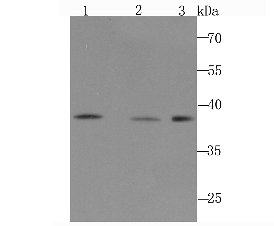
Fig1: Western blot analysis of DNA Polymerase beta on different cell lysate using anti-DNA Polymerase beta antibody at 1/1,000 dilution. Positive control: Lane1: PC-12 Lane2: NIH-3T3 Lane3: A431Application
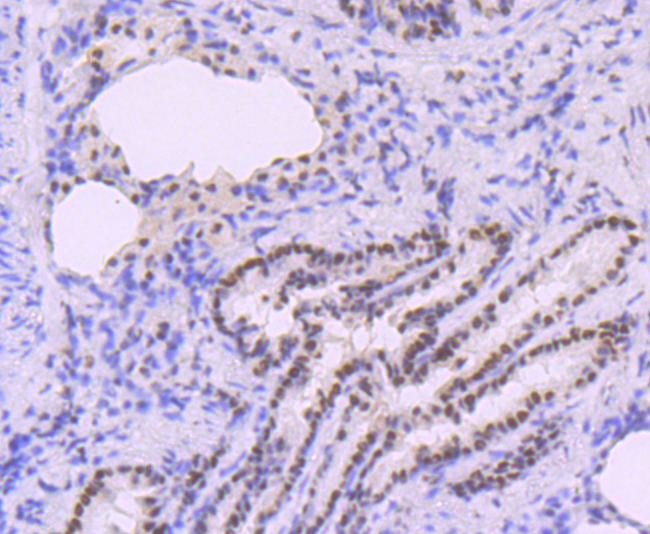
Fig2: Immunohistochemical analysis of paraffin-embedded rat lung tissue using anti-DNA Polymerase beta antibody. Counter stained with hematoxylin.Application
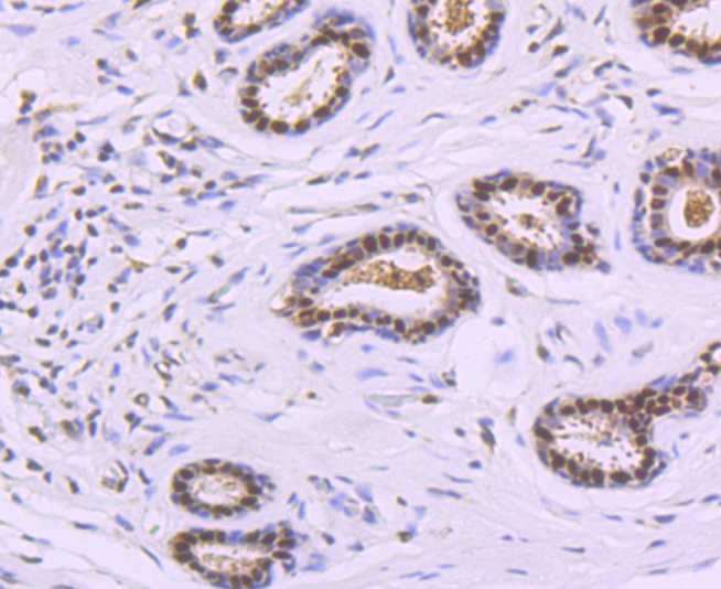
Fig3: Immunohistochemical analysis of paraffin-embedded human breast cancer tissue using anti-DNA Polymerase beta antibody. Counter stained with hematoxylin.Application
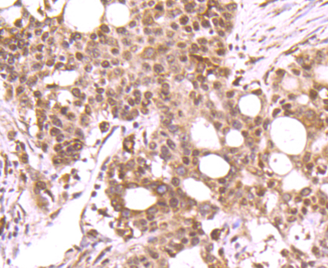
Fig4: Immunohistochemical analysis of paraffin-embedded human stomach cancer tissue using anti-DNA Polymerase beta antibody. Counter stained with hematoxylin.Application
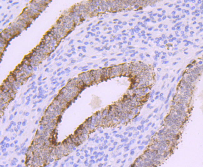
Fig5: Immunohistochemical analysis of paraffin-embedded human uterus tissue using anti-DNA Polymerase beta antibody. Counter stained with hematoxylin.Application
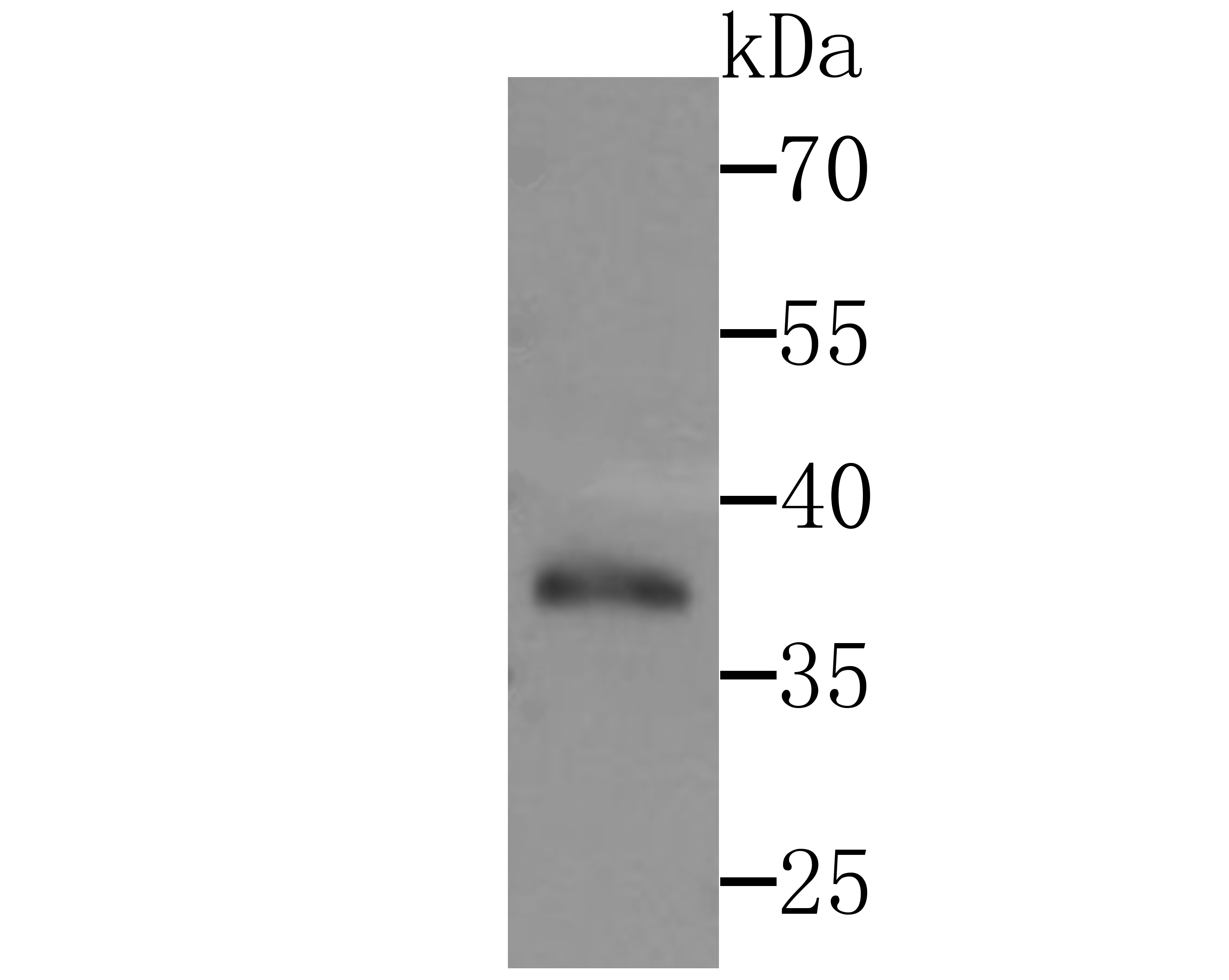
Fig6: Western blot analysis of DNA Polymerase beta on Zebrafish tissue lysates using anti-DNA Polymerase beta antibody at 1/200 dilution.Application
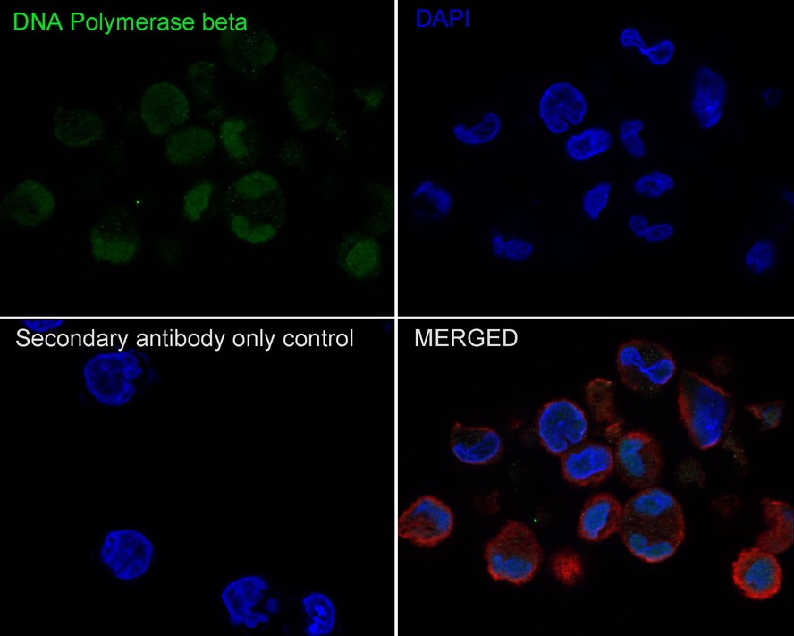
Fig7:Immunocytochemistry analysis of K-562 cells labeling DNA Polymerase beta with Rabbit anti-DNA Polymerase beta antibody at 1/100 dilution. Cells were fixed in 4% paraformaldehyde for 10 minutes at 37 ℃, permeabilized with 0.05% Triton X-100 in PBS for 20 minutes, and then blocked with 2% negative goat serum for 30 minutes at room temperature. Cells were then incubated with Rabbit anti-DNA Polymerase beta antibody at 1/100 dilution in 2% negative goat serum overnight at 4 ℃. Goat Anti-Rabbit IgG H&L (488) was used as the secondary antibody at 1/1,000 dilution. Nuclear DNA was labelled in blue with DAPI. Beta tubulin (red) was stained at 1/200 dilution overnight at +4℃. Goat Anti-Mouse IgG H&L (594) was used as the secondary antibody at 1/1,000 dilution.| Product Name | DNA Polymerase beta Recombinant Rabbit Monoclonal Antibody |
|---|---|
| Antibody Type | Primary Antibodies |
| Immunogen | Synthetic peptide within Human DNA Polymerase beta aa 286-335 / 335. |
| Clonality | Monoclonal |
|---|---|
| Isotype | IgG |
| Host Species | Recombinant rabbit |
| Tested Applications | ICC/IFIHCWB |
| WB:1:500 IHC:1:50-1:200 ICC/IF:1:20-1:50 |
|
| Species Reactivity | HumanMouseRatZebrafish |
| Concentration | 1mg/ml |
| Purification | Protein A |
| Alternative Names | DNA directed DNA polymerase beta antibody DNA pol beta antibody DNA polymerase beta antibody DNA polymerase beta subunit antibody DPOLB_HUMAN antibody MGC125976 antibody Pol B antibody Pol beta antibody POLB antibody Polymerase (DNA directed) beta antibody |
|---|---|
| Molecular Weight(MW) | 38 kDa |
| Cellular Localization | Nucleus, Cytoplasm. |
Application
Fig1: Western blot analysis of DNA Polymerase beta on different cell lysate using anti-DNA Polymerase beta antibody at 1/1,000 dilution. Positive control: Lane1: PC-12 Lane2: NIH-3T3 Lane3: A431
Application
Fig2: Immunohistochemical analysis of paraffin-embedded rat lung tissue using anti-DNA Polymerase beta antibody. Counter stained with hematoxylin.
Application
Fig3: Immunohistochemical analysis of paraffin-embedded human breast cancer tissue using anti-DNA Polymerase beta antibody. Counter stained with hematoxylin.
Application
Fig4: Immunohistochemical analysis of paraffin-embedded human stomach cancer tissue using anti-DNA Polymerase beta antibody. Counter stained with hematoxylin.
Application
Fig5: Immunohistochemical analysis of paraffin-embedded human uterus tissue using anti-DNA Polymerase beta antibody. Counter stained with hematoxylin.
Application
Fig6: Western blot analysis of DNA Polymerase beta on Zebrafish tissue lysates using anti-DNA Polymerase beta antibody at 1/200 dilution.
Application
Fig7:Immunocytochemistry analysis of K-562 cells labeling DNA Polymerase beta with Rabbit anti-DNA Polymerase beta antibody at 1/100 dilution. Cells were fixed in 4% paraformaldehyde for 10 minutes at 37 ℃, permeabilized with 0.05% Triton X-100 in PBS for 20 minutes, and then blocked with 2% negative goat serum for 30 minutes at room temperature. Cells were then incubated with Rabbit anti-DNA Polymerase beta antibody at 1/100 dilution in 2% negative goat serum overnight at 4 ℃. Goat Anti-Rabbit IgG H&L (488) was used as the secondary antibody at 1/1,000 dilution. Nuclear DNA was labelled in blue with DAPI. Beta tubulin (red) was stained at 1/200 dilution overnight at +4℃. Goat Anti-Mouse IgG H&L (594) was used as the secondary antibody at 1/1,000 dilution.| Application Notes | WB:1:500 IHC:1:50-1:200 ICC/IF:1:20-1:50 |
|---|
| Form | Liquid |
|---|---|
| Storage Instructions | Store at +4℃ after thawing. Aliquot store at -20℃ or -80℃. Avoid repeated freeze / thaw cycles. |
| Storage Buffer | 1*TBS (pH7.4), 1%BSA, 40%Glycerol. Preservative: 0.05% Sodium Azide. |


