Application
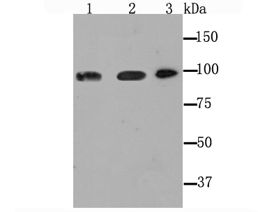
Fig1: Western blot analysis of Dynamin 2 on different cell lysate using anti-Dynamin 2 antibody at 1/1,000 dilution. Positive control: Lane1: PC-12 Lane2: Mouse thymus Lane3: HelaApplication
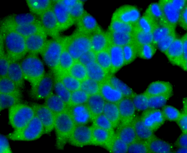
Fig2: ICC staining Dynamin 2 in 293T cells (green). The nuclear counter stain is DAPI (blue). Cells were fixed in paraformaldehyde, permeabilised with 0.25% Triton X100/PBS.Application
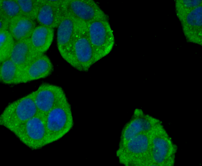
Fig3: ICC staining Dynamin 2 in Hela cells (green). The nuclear counter stain is DAPI (blue). Cells were fixed in paraformaldehyde, permeabilised with 0.25% Triton X100/PBS.Application
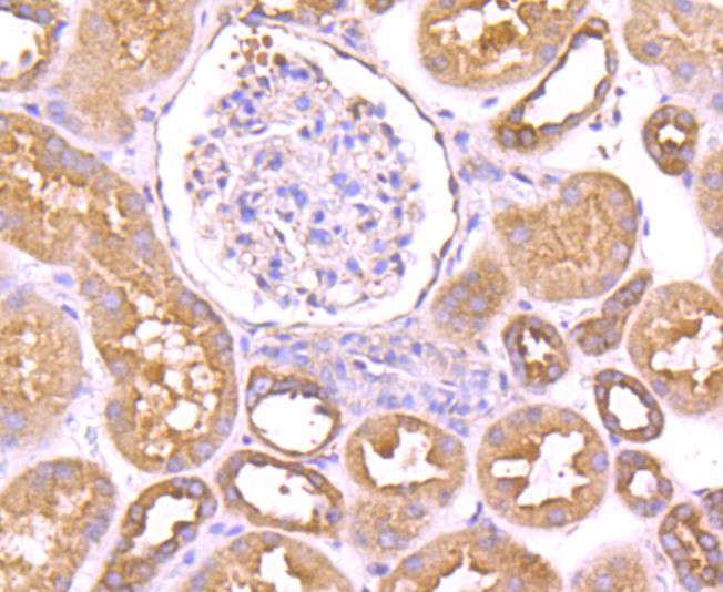
Fig4: Immunohistochemical analysis of paraffin-embedded human kidney tissue using anti-Dynamin 2 antibody. Counter stained with hematoxylin.Application
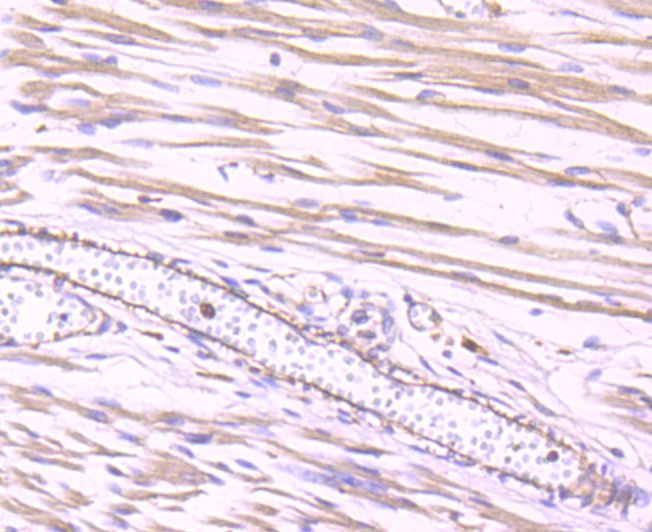
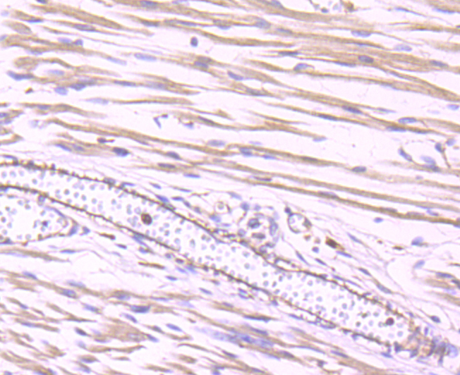
Fig5: Immunohistochemical analysis of paraffin-embedded human fetal skeletal muscle tissue using anti-Dynamin 2 antibody. Counter stained with hematoxylin.Application
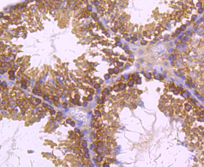
Fig6: Immunohistochemical analysis of paraffin-embedded mouse testes tissue using anti-Dynamin 2 antibody. Counter stained with hematoxylin.Application
Fig7: Immunohistochemical analysis of paraffin-embedded mouse colon tissue using anti-Dynamin 2 antibody. Counter stained with hematoxylin.Application
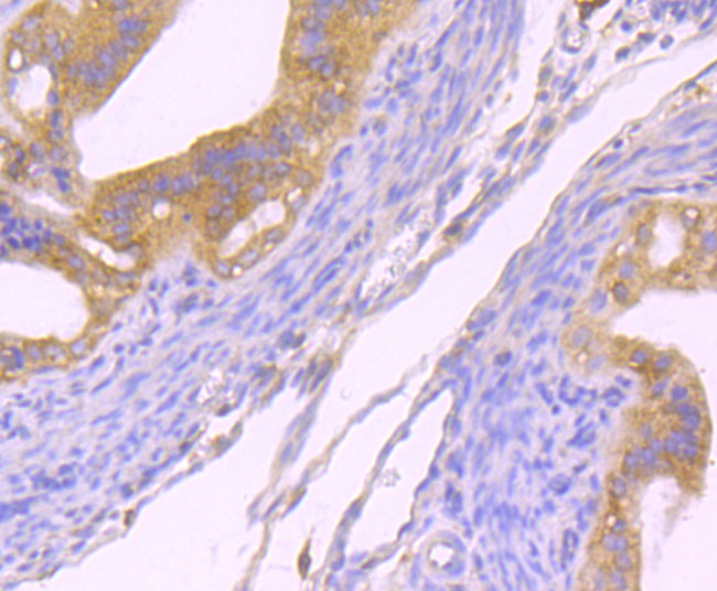
Fig8: Immunohistochemical analysis of paraffin-embedded mouse fallopian tube tissue using anti-Dynamin 2 antibody. Counter stained with hematoxylin.Application
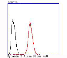
Fig9: Flow cytometric analysis of Hela cells with Dynamin 2 antibody at 1/100 dilution (red) compared with an unlabelled control (cells without incubation with primary antibody; black).| Product Name | Dynamin 2 [JM61-38] |
|---|---|
| Antibody Type | Primary Antibodies |
| Product description | Members of the dynamin family, including Dynamin I and Dynamin II, are GPTase, microtubule-associated proteins which are involved in endocytosis, synaptic transmission and neurogenesis. Dynamin I is localized to the central nervous system, while Dynamin II exhibits ubiquitous distribution with highest expression found in testis. Both dynamin proteins contain SH3 and proline-rich domains that mediate interactions between the dynamins and effectors of their GTPase activity. The interactions with these effectors, which include microtubules, acidic phospholipids and SH3 domain-containing proteins, are required for rapid endocytosis. Dynamin I appears to be recruited to clathrin coated pits by SH3 domain interaction with amphiphysin, a protein highly expressed in brain. |
| Immunogen | Recombinant protein |
| Clonality | Monoclonal |
|---|---|
| Isotype | IgG |
| Host Species | Recombinant rabbit |
| Tested Applications | WB,ICC/IF,IHC,FC |
| Species Reactivity | Human |
| Concentration | 1mg/ml |
| Purification | Unpurified |
| Alternative Names | CMT2M antibody CMTDI1 antibody CMTDIB antibody DI CMTB antibody Dnm2 antibody DYN II antibody DYN2 antibody DYN2_HUMAN antibody Dynamin II antibody Dynamin-2 antibody Dynamin2 antibody DynaminII antibody DYNII antibody |
|---|---|
| Molecular Weight(MW) | 98 kDa |
| Cellular Localization | Cytoplasm. |
Application
Fig1: Western blot analysis of Dynamin 2 on different cell lysate using anti-Dynamin 2 antibody at 1/1,000 dilution. Positive control: Lane1: PC-12 Lane2: Mouse thymus Lane3: Hela
Application
Fig2: ICC staining Dynamin 2 in 293T cells (green). The nuclear counter stain is DAPI (blue). Cells were fixed in paraformaldehyde, permeabilised with 0.25% Triton X100/PBS.
Application
Fig3: ICC staining Dynamin 2 in Hela cells (green). The nuclear counter stain is DAPI (blue). Cells were fixed in paraformaldehyde, permeabilised with 0.25% Triton X100/PBS.
Application
Fig4: Immunohistochemical analysis of paraffin-embedded human kidney tissue using anti-Dynamin 2 antibody. Counter stained with hematoxylin.
Application
Fig5: Immunohistochemical analysis of paraffin-embedded human fetal skeletal muscle tissue using anti-Dynamin 2 antibody. Counter stained with hematoxylin.
Application
Fig6: Immunohistochemical analysis of paraffin-embedded mouse testes tissue using anti-Dynamin 2 antibody. Counter stained with hematoxylin.
Application
Fig7: Immunohistochemical analysis of paraffin-embedded mouse colon tissue using anti-Dynamin 2 antibody. Counter stained with hematoxylin.
Application
Fig8: Immunohistochemical analysis of paraffin-embedded mouse fallopian tube tissue using anti-Dynamin 2 antibody. Counter stained with hematoxylin.
Application
Fig9: Flow cytometric analysis of Hela cells with Dynamin 2 antibody at 1/100 dilution (red) compared with an unlabelled control (cells without incubation with primary antibody; black).| Positive Control | PC-12, mouse thymus tissue, Hela, 293T, human kidney tissue, human fetal skeletal muscle tissue, mouse testes tissue, mouse colon tissue, mouse fallopian tube tissue. |
|---|
| Form | Liquid |
|---|---|
| Storage Instructions | Store at +4℃ after thawing. Aliquot store at -20℃ or -80℃. Avoid repeated freeze / thaw cycles. |
| Storage Buffer | 1*TBS (pH7.4), 1%BSA, 40%Glycerol. Preservative: 0.05% Sodium Azide. |


