WB
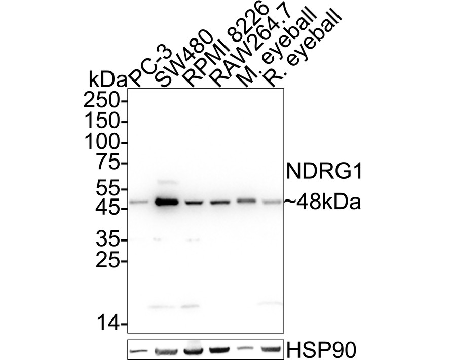
Western blot analysis of NDRG1 on different lysates with Rabbit anti-NDRG1 antibody at 1/1,000 dilution. Lane 1: PC-3 cell lysate (20 µg/Lane), Lane 2: SW480 cell lysate (20 µg/Lane), Lane 3: RPMI 8226 cell lysate (20 µg/Lane), Lane 4: RAW264.7 cell lysate (20 µg/Lane), Lane 5: Mouse eyeball tissue lysate (40 µg/Lane), Lane 6: Rat eyeball tissue lysate (40 µg/Lane), Lysates/proteins at 10 µg/Lane. Exposure time: 6 seconds; 4-20% SDS-PAGE gel. Proteins were transferred to a PVDF membrane and blocked with 5% NFDM/TBST for 1 hour at room temperature. The primary antibody at 1/1,000 dilution was used in 5% NFDM/TBST at 4℃ overnight. Goat Anti-Rabbit IgG - HRP Secondary Antibody at 1/50,000 dilution was used for 1 hour at room temperature.ICC/IF
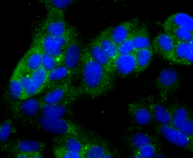
ICC staining NDRG1 in MCF-7 cells (green). The nuclear counter stain is DAPI (blue). Cells were fixed in paraformaldehyde, permeabilised with 0.25% Triton X100/PBS.ICC/IF
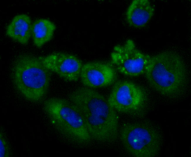
ICC staining NDRG1 in A431 cells (green). The nuclear counter stain is DAPI (blue). Cells were fixed in paraformaldehyde, permeabilised with 0.25% Triton X100/PBS.IHC
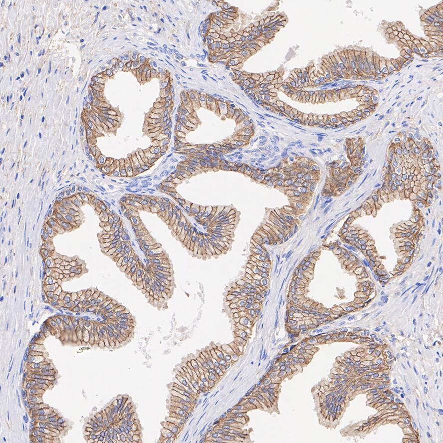
Immunohistochemical analysis of paraffin-embedded human prostate tissue with Rabbit anti-NDRG1 antibody at 1/1,000 dilution. The section was pre-treated using heat mediated antigen retrieval with Tris-EDTA buffer (pH 9.0) for 20 minutes. The tissues were blocked in 1% BSA for 20 minutes at room temperature, washed with ddH2O and PBS, and then probed with the primary antibody at 1/1,000 dilution for 1 hour at room temperature. The detection was performed using an HRP conjugated compact polymer system. DAB was used as the chromogen. Tissues were counterstained with hematoxylin and mounted with DPX.IHC
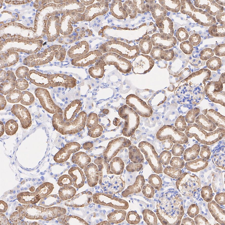
Immunohistochemical analysis of paraffin-embedded mouse kidney tissue with Rabbit anti-NDRG1 antibody at 1/1,000 dilution. The section was pre-treated using heat mediated antigen retrieval with Tris-EDTA buffer (pH 9.0) for 20 minutes. The tissues were blocked in 1% BSA for 20 minutes at room temperature, washed with ddH2O and PBS, and then probed with the primary antibody at 1/1,000 dilution for 1 hour at room temperature. The detection was performed using an HRP conjugated compact polymer system. DAB was used as the chromogen. Tissues were counterstained with hematoxylin and mounted with DPX.Application
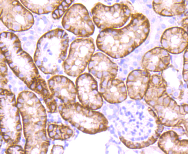
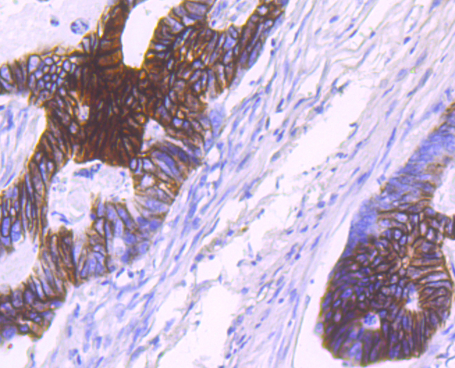
Immunohistochemical analysis of paraffin-embedded mouse kidney tissue using anti-NDRG1 antibody. Counter stained with hematoxylin.Immunohistochemical analysis of paraffin-embedded human colon cancer tissue using anti-NDRG1 antibody. Counter stained with hematoxylin.
| Product Name | NDRG1 Recombinant Rabbit Monoclonal Antibody |
|---|---|
| Antibody Type | Primary Antibodies |
| Product description | The N-myc downstream regulated gene (NDRG) family is comprised of four members, NDRG1 (also designated Drg1, RTP, rit42, Cap43 and Ndr1), NDRG2, NDRG3 and NDRG4, which share 57-65% homology. The NDRG1 gene, which maps to human chromosome 8q24.22, is evolutionarily conserved and is similarly regulated in humans, mice and rats . Like NDRG2 and NDRG3, NDRG1 is ubiquitously expressed, but it is expressed most prominently in placental membranes and prostate, kidney, small intestine and ovary tissue. NDRG1 gene expression is induced by several compounds, including nickel, and produces a protein involved in stress responses, hormone responses, cell growth and differentiation. The gene encoding NDRG3 maps to human chromosome 20q11.23 and is predominantly expressed in testis, prostate and ovary, which suggests it may play a role in spermatogenesis. |
| Immunogen | Synthetic peptide within Human NDRG1 aa 345-394 / 394. |
| Clonality | Monoclonal |
|---|---|
| Isotype | IgG |
| Host Species | Recombinant rabbit |
| Tested Applications | ICC/IFIHCWB |
| WB:1:500-1:2000 IHC:1:200-1:1000 ICC/IF:1:50-1:200 |
|
| Species Reactivity | HumanMouseRat |
| Concentration | 1mg/ml |
| Purification | Protein A |
| Alternative Names | 42 kDa antibody Anti GC4 antibody cap43 antibody cmt4d antibody Differentiation related gene1 protein antibody Differentiation-related gene 1 protein antibody Drg 1 antibody DRG-1 antibody drg1 antibody gc4 antibody GC4 antibody antibody hmsnl antibody Human mRNA for RTP complete cds antibody N myc downstream regulated gene 1 antibody N myc downstream regulated gene 1 protein antibody N-myc downstream-regulated gene 1 protein antibody Ndr 1 antibody ndr1 antibody NDRG 1 antibody Ndrg1 antibody NDRG1 protein antibody NDRG1_HUMAN antibody Nickel specific induction protein antibody Nickel specific induction protein Cap43 antibody Nickel-specific induction protein Cap43 antibody nmsl antibody Nmyc downstream regulated antibody Nmyc downstream regulated gene1 antibody Nmyc downstream regulated gene1 protein antibody Protein NDRG1 antibody Protein regulated by oxygen 1 antibody Protein regulated by oxygen1 antibody Proxy1 antibody Reduced in tumor antibody Reducin antibody Reducing agents and tunicamycin responsive protein antibody Reducing agents and tunicamycin-responsive protein antibody Rit42 antibody RTP antibody targ1 antibody TDD5 antibody tdds antibody Tunicamycin responsive protein antibody |
|---|---|
| Molecular Weight(MW) | 43 kDa(Observed band size: 48 kDa) |
| Cellular Localization | Cytoplasm. Nucleus. Cell membrane. |
WB
Western blot analysis of NDRG1 on different lysates with Rabbit anti-NDRG1 antibody at 1/1,000 dilution. Lane 1: PC-3 cell lysate (20 µg/Lane), Lane 2: SW480 cell lysate (20 µg/Lane), Lane 3: RPMI 8226 cell lysate (20 µg/Lane), Lane 4: RAW264.7 cell lysate (20 µg/Lane), Lane 5: Mouse eyeball tissue lysate (40 µg/Lane), Lane 6: Rat eyeball tissue lysate (40 µg/Lane), Lysates/proteins at 10 µg/Lane. Exposure time: 6 seconds; 4-20% SDS-PAGE gel. Proteins were transferred to a PVDF membrane and blocked with 5% NFDM/TBST for 1 hour at room temperature. The primary antibody at 1/1,000 dilution was used in 5% NFDM/TBST at 4℃ overnight. Goat Anti-Rabbit IgG - HRP Secondary Antibody at 1/50,000 dilution was used for 1 hour at room temperature.
ICC/IF
ICC staining NDRG1 in MCF-7 cells (green). The nuclear counter stain is DAPI (blue). Cells were fixed in paraformaldehyde, permeabilised with 0.25% Triton X100/PBS.
ICC/IF
ICC staining NDRG1 in A431 cells (green). The nuclear counter stain is DAPI (blue). Cells were fixed in paraformaldehyde, permeabilised with 0.25% Triton X100/PBS.
IHC
Immunohistochemical analysis of paraffin-embedded human prostate tissue with Rabbit anti-NDRG1 antibody at 1/1,000 dilution. The section was pre-treated using heat mediated antigen retrieval with Tris-EDTA buffer (pH 9.0) for 20 minutes. The tissues were blocked in 1% BSA for 20 minutes at room temperature, washed with ddH2O and PBS, and then probed with the primary antibody at 1/1,000 dilution for 1 hour at room temperature. The detection was performed using an HRP conjugated compact polymer system. DAB was used as the chromogen. Tissues were counterstained with hematoxylin and mounted with DPX.
IHC
Immunohistochemical analysis of paraffin-embedded mouse kidney tissue with Rabbit anti-NDRG1 antibody at 1/1,000 dilution. The section was pre-treated using heat mediated antigen retrieval with Tris-EDTA buffer (pH 9.0) for 20 minutes. The tissues were blocked in 1% BSA for 20 minutes at room temperature, washed with ddH2O and PBS, and then probed with the primary antibody at 1/1,000 dilution for 1 hour at room temperature. The detection was performed using an HRP conjugated compact polymer system. DAB was used as the chromogen. Tissues were counterstained with hematoxylin and mounted with DPX.
Application
Immunohistochemical analysis of paraffin-embedded mouse kidney tissue using anti-NDRG1 antibody. Counter stained with hematoxylin.
Immunohistochemical analysis of paraffin-embedded human colon cancer tissue using anti-NDRG1 antibody. Counter stained with hematoxylin.
| Application Notes | WB:1:500-1:2000 IHC:1:200-1:1000 ICC/IF:1:50-1:200 |
|---|
| Form | Liquid |
|---|---|
| Storage Instructions | Store at +4℃ after thawing. Aliquot store at -20℃ or -80℃. Avoid repeated freeze / thaw cycles. |
| Storage Buffer | 1*TBS (pH7.4), 1%BSA, 40%Glycerol. Preservative: 0.05% Sodium Azide. |


