WB
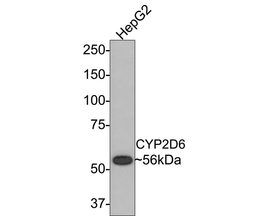
Western blot analysis of CYP2D6 on HepG2 cell lysates with Rabbit anti-CYP2D6 antibody at 1/500 dilution. Lysates/proteins at 10 µg/Lane. Exposure time: 2 minutes; 8% SDS-PAGE gel. Proteins were transferred to a PVDF membrane and blocked with 5% NFDM/TBST for 1 hour at room temperature. The primary antibody at 1/500 dilution was used in 5% NFDM/TBST at room temperature for 2 hours. Goat Anti-Rabbit IgG - HRP Secondary Antibody at 1:300,000 dilution was used for 1 hour at room temperature.ICC/IF
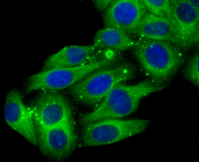
ICC staining of CYP2D6 in HepG2 cells (green). Formalin fixed cells were permeabilized with 0.1% Triton X-100 in TBS for 10 minutes at room temperature and blocked with 10% negative goat serum for 15 minutes at room temperature. Cells were probed with the primary antibody (1/50) for 1 hour at room temperature, washed with PBS. Alexa Fluor®488 conjugate-Goat anti-Rabbit IgG was used as the secondary antibody at 1/1,000 dilution. The nuclear counter stain is DAPI (blue).ICC/IF
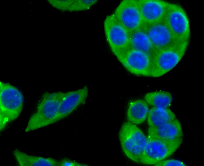
ICC staining of CYP2D6 in SW480 cells (green). Formalin fixed cells were permeabilized with 0.1% Triton X-100 in TBS for 10 minutes at room temperature and blocked with 10% negative goat serum for 15 minutes at room temperature. Cells were probed with the primary antibody (1/50) for 1 hour at room temperature, washed with PBS. Alexa Fluor®488 conjugate-Goat anti-Rabbit IgG was used as the secondary antibody at 1/1,000 dilution. The nuclear counter stain is DAPI (blue).IHC
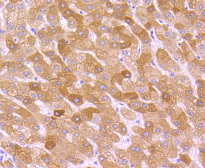
Immunohistochemical analysis of paraffin-embedded human liver tissue using anti-CYP2D6 antibody. The section was pre-treated using heat mediated antigen retrieval with Tris-EDTA buffer (pH 9.0) for 20 minutes.The tissues were blocked in 1% BSA for 30 minutes at room temperature, washed with ddH2O and PBS, and then probed with the primary antibody (1/50) for 30 minutes at room temperature. The detection was performed using an HRP conjugated compact polymer system. DAB was used as the chromogen. Tissues were counterstained with hematoxylin and mounted with DPX.IHC
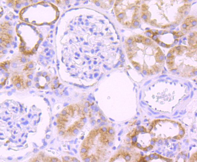
Immunohistochemical analysis of paraffin-embedded human kidney tissue using anti-CYP2D6 antibody. The section was pre-treated using heat mediated antigen retrieval with Tris-EDTA buffer (pH 9.0) for 20 minutes.The tissues were blocked in 1% BSA for 30 minutes at room temperature, washed with ddH2O and PBS, and then probed with the primary antibody (1/50) for 30 minutes at room temperature. The detection was performed using an HRP conjugated compact polymer system. DAB was used as the chromogen. Tissues were counterstained with hematoxylin and mounted with DPX.FC
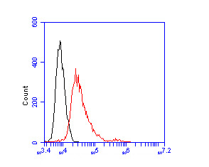
Flow cytometric analysis of CYP2D6 was done on SW480 cells. The cells were fixed, permeabilized and stained with the primary antibody (1/50) (red). After incubation of the primary antibody at room temperature for an hour, the cells were stained with a Alexa Fluor®488 conjugate-Goat anti-Rabbit IgG Secondary antibody at 1/1,000 dilution for 30 minutes.Unlabelled sample was used as a control (cells without incubation with primary antibody; black).| Product Name | CYP2D6 Recombinant Rabbit Monoclonal Antibody |
|---|---|
| Antibody Type | Primary Antibodies |
| Product description | The P450II family comprises at least five subfamilies, designated A through E by the system of nomenclature recommended by an international committee. The P450IID subfamily comprises at least two genes in the rat, one of which is highly specific for debrisoquine 4-hydroxylase activity. An association of this gene with lung cancer has been found. Enhanced CYP2D6 activity has been related to malignancies of the bladder, liver, pharynx and stomach, and especially to cigarette-smoking-induced lung cancer. The data suggests that enhanced CYP2D6-mediated metabolism of one or more dietary and other environmental agents, to form a reactive intermediate, plays a role in cancer initiation and/or promotion in various tissues. CYP2D6 polymorphism, which is responsible for the variation in metabolism of debrisoquine 4-hydroxylase, is important in the metabolism of more than 30 drugs and environmental chemicals, including as much as 20% of all commonly prescribed drugs. The gene which encodes CYP2D6 maps to human chromosome 22q13.1. |
| Immunogen | Recombinant protein within Human CYP2D6 aa 301-497 / 497. |
| Clonality | Monoclonal |
|---|---|
| Isotype | IgG |
| Host Species | Recombinant rabbit |
| Tested Applications | FCICC/IFIHCWB |
| WB:1:500-1:2,000 ICC/IF:1:50-1:200 IHC:1:50-1:200 FC:1:50-1:100 |
|
| Species Reactivity | Human |
| Concentration | 1mg/ml |
| Purification | Protein A |
| Alternative Names | CPD6 antibody CYP2D antibody CYP2D6 antibody CYP2D7AP antibody CYP2D7BP antibody CYP2D7P2 antibody CYP2D8P2 antibody CYP2DL1 antibody CYPIID6 antibody Cytochrome P450 DB1 antibody Cytochrome P450 family 2 subfamily D member 6 antibody Cytochrome P450 family 2 subfamily D polypeptide 6 antibody Debrisoquine 4 hydroxylase antibody Flavoprotein linked monooxygenase antibody Microsomal monooxygenase antibody P450 DB1 antibody P450C2D antibody P450DB1 antibody Xenobiotic monooxygenase antibody |
|---|---|
| Molecular Weight(MW) | 56 kDa |
| Cellular Localization | Endoplasmic reticulum membrane; Peripheral membrane protein. Microsome membrane; Peripheral membrane protein. |
| SwissProt ID | P10635 |
|---|
WB
Western blot analysis of CYP2D6 on HepG2 cell lysates with Rabbit anti-CYP2D6 antibody at 1/500 dilution. Lysates/proteins at 10 µg/Lane. Exposure time: 2 minutes; 8% SDS-PAGE gel. Proteins were transferred to a PVDF membrane and blocked with 5% NFDM/TBST for 1 hour at room temperature. The primary antibody at 1/500 dilution was used in 5% NFDM/TBST at room temperature for 2 hours. Goat Anti-Rabbit IgG - HRP Secondary Antibody at 1:300,000 dilution was used for 1 hour at room temperature.
ICC/IF
ICC staining of CYP2D6 in HepG2 cells (green). Formalin fixed cells were permeabilized with 0.1% Triton X-100 in TBS for 10 minutes at room temperature and blocked with 10% negative goat serum for 15 minutes at room temperature. Cells were probed with the primary antibody (1/50) for 1 hour at room temperature, washed with PBS. Alexa Fluor®488 conjugate-Goat anti-Rabbit IgG was used as the secondary antibody at 1/1,000 dilution. The nuclear counter stain is DAPI (blue).
ICC/IF
ICC staining of CYP2D6 in SW480 cells (green). Formalin fixed cells were permeabilized with 0.1% Triton X-100 in TBS for 10 minutes at room temperature and blocked with 10% negative goat serum for 15 minutes at room temperature. Cells were probed with the primary antibody (1/50) for 1 hour at room temperature, washed with PBS. Alexa Fluor®488 conjugate-Goat anti-Rabbit IgG was used as the secondary antibody at 1/1,000 dilution. The nuclear counter stain is DAPI (blue).
IHC
Immunohistochemical analysis of paraffin-embedded human liver tissue using anti-CYP2D6 antibody. The section was pre-treated using heat mediated antigen retrieval with Tris-EDTA buffer (pH 9.0) for 20 minutes.The tissues were blocked in 1% BSA for 30 minutes at room temperature, washed with ddH2O and PBS, and then probed with the primary antibody (1/50) for 30 minutes at room temperature. The detection was performed using an HRP conjugated compact polymer system. DAB was used as the chromogen. Tissues were counterstained with hematoxylin and mounted with DPX.
IHC
Immunohistochemical analysis of paraffin-embedded human kidney tissue using anti-CYP2D6 antibody. The section was pre-treated using heat mediated antigen retrieval with Tris-EDTA buffer (pH 9.0) for 20 minutes.The tissues were blocked in 1% BSA for 30 minutes at room temperature, washed with ddH2O and PBS, and then probed with the primary antibody (1/50) for 30 minutes at room temperature. The detection was performed using an HRP conjugated compact polymer system. DAB was used as the chromogen. Tissues were counterstained with hematoxylin and mounted with DPX.
FC
Flow cytometric analysis of CYP2D6 was done on SW480 cells. The cells were fixed, permeabilized and stained with the primary antibody (1/50) (red). After incubation of the primary antibody at room temperature for an hour, the cells were stained with a Alexa Fluor®488 conjugate-Goat anti-Rabbit IgG Secondary antibody at 1/1,000 dilution for 30 minutes.Unlabelled sample was used as a control (cells without incubation with primary antibody; black).| Application Notes | WB:1:500-1:2,000 ICC/IF:1:50-1:200 IHC:1:50-1:200 FC:1:50-1:100 |
|---|
| Form | Liquid |
|---|---|
| Storage Instructions | Store at +4℃ after thawing. Aliquot store at -20℃ or -80℃. Avoid repeated freeze / thaw cycles. |
| Storage Buffer | 1*TBS (pH7.4), 1%BSA, 40%Glycerol. Preservative: 0.05% Sodium Azide. |


