WB
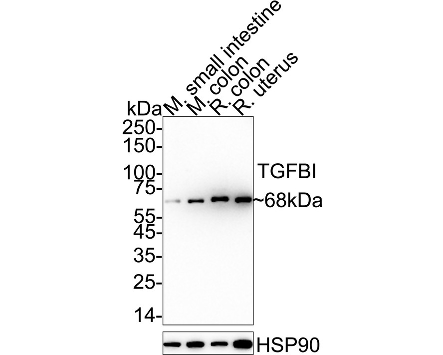
Western blot analysis of TGFBI on different lysates with Rabbit anti-TGFBI antibody at 1/1,000 dilution. Lane 1: Mouse small intestine tissue lysate (20 µg/Lane), Lane 2: Mouse colon tissue lysate (20 µg/Lane), Lane 3: Rat colon tissue lysate (20 µg/Lane), Lane 4: Rat uterus tissue lysate (20 µg/Lane), Exposure time: 3 minutes; 4-20% SDS-PAGE gel. Proteins were transferred to a PVDF membrane and blocked with 5% NFDM/TBST for 1 hour at room temperature. The primary antibody at 1/1,000 dilution was used in 5% NFDM/TBST at 4℃ overnight. Goat Anti-Rabbit IgG - HRP Secondary Antibody at 1/50,000 dilution was used for 1 hour at room temperature.IHC
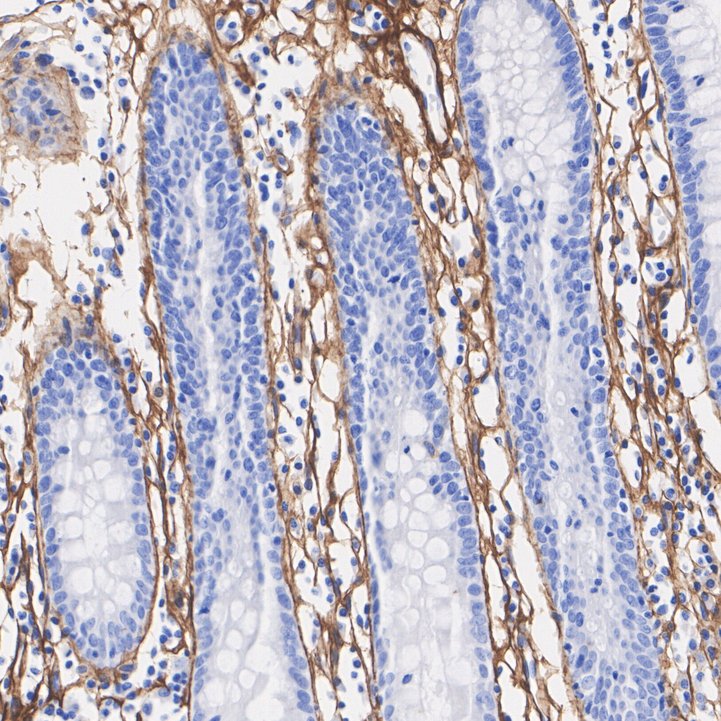
Immunohistochemical analysis of paraffin-embedded human colon tissue with Rabbit anti-TGFBI antibody at 1/200 dilution. The section was pre-treated using heat mediated antigen retrieval with Tris-EDTA buffer (pH 9.0) for 20 minutes. The tissues were blocked in 1% BSA for 20 minutes at room temperature, washed with ddH2O and PBS, and then probed with the primary antibody at 1/200 dilution for 1 hour at room temperature. The detection was performed using an HRP conjugated compact polymer system. DAB was used as the chromogen. Tissues were counterstained with hematoxylin and mounted with DPX.IHC
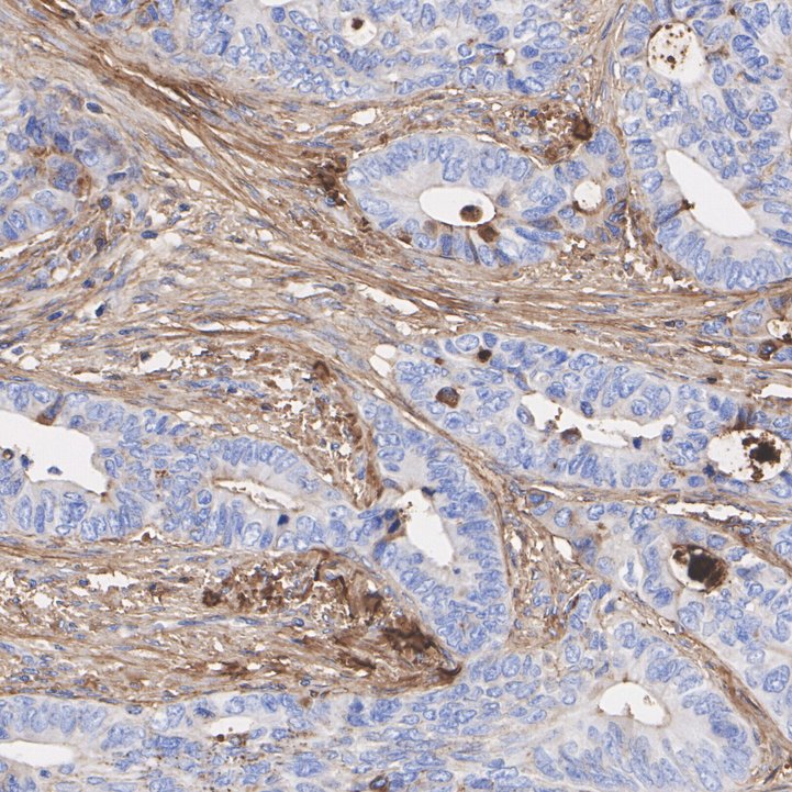
Immunohistochemical analysis of paraffin-embedded human colon cancer tissue with Rabbit anti-TGFBI antibody at 1/200 dilution. The section was pre-treated using heat mediated antigen retrieval with Tris-EDTA buffer (pH 9.0) for 20 minutes. The tissues were blocked in 1% BSA for 20 minutes at room temperature, washed with ddH2O and PBS, and then probed with the primary antibody at 1/200 dilution for 1 hour at room temperature. The detection was performed using an HRP conjugated compact polymer system. DAB was used as the chromogen. Tissues were counterstained with hematoxylin and mounted with DPX.| Product Name | TGFBI Recombinant Rabbit Monoclonal Antibody |
|---|---|
| Antibody Type | Primary Antibodies |
| Product description | Binds to type I, II, and IV collagens. This adhesion protein may play an important role in cell-collagen interactions. In cartilage, may be involved in endochondral bone formation.Highly expressed in the corneal epithelium. |
| Immunogen | Synthetic peptide within Human TGFBI aa 93-142 / 683. |
| Clonality | Monoclonal |
|---|---|
| Isotype | IgG |
| Host Species | Recombinant rabbit |
| Tested Applications | IHCWB |
| WB:1:1000 IHC:1:200 |
|
| Species Reactivity | HumanMouseRat |
| Concentration | 1mg/ml |
| Purification | Protein A |
| Alternative Names | RGD containing collagen associated protein antibody
AI181842 antibody
AI747162 antibody
Beta ig antibody
Beta ig h3 antibody
Beta ig-h3 antibody
BGH3_HUMAN antibody
Big h3 antibody
BIGH3 antibody
CDB1 antibody
CDG2 antibody
CDGG1 antibody
CSD antibody
CSD1 antibody
CSD2 antibody
CSD3 antibody
EBMD antibody
Kerato epithelin antibody
Kerato-epithelin antibody
LCD1 antibody
MGC150270 antibody
RGD CAP antibody
RGD-CAP antibody
RGD-containing collagen-associated protein antibody
TGFBI antibody
TGFBI transforming growth factor beta induced 68kDa antibody Transforming growth factor beta induced protein ig h3 antibody Transforming growth factor-beta-induced protein ig-h3 antibody |
|---|---|
| Molecular Weight(MW) | 75kDa(Observed band size: 68kDa) |
| Cellular Localization | Secreted, extracellular space, extracellular matrix. |
WB
Western blot analysis of TGFBI on different lysates with Rabbit anti-TGFBI antibody at 1/1,000 dilution. Lane 1: Mouse small intestine tissue lysate (20 µg/Lane), Lane 2: Mouse colon tissue lysate (20 µg/Lane), Lane 3: Rat colon tissue lysate (20 µg/Lane), Lane 4: Rat uterus tissue lysate (20 µg/Lane), Exposure time: 3 minutes; 4-20% SDS-PAGE gel. Proteins were transferred to a PVDF membrane and blocked with 5% NFDM/TBST for 1 hour at room temperature. The primary antibody at 1/1,000 dilution was used in 5% NFDM/TBST at 4℃ overnight. Goat Anti-Rabbit IgG - HRP Secondary Antibody at 1/50,000 dilution was used for 1 hour at room temperature.
IHC
Immunohistochemical analysis of paraffin-embedded human colon tissue with Rabbit anti-TGFBI antibody at 1/200 dilution. The section was pre-treated using heat mediated antigen retrieval with Tris-EDTA buffer (pH 9.0) for 20 minutes. The tissues were blocked in 1% BSA for 20 minutes at room temperature, washed with ddH2O and PBS, and then probed with the primary antibody at 1/200 dilution for 1 hour at room temperature. The detection was performed using an HRP conjugated compact polymer system. DAB was used as the chromogen. Tissues were counterstained with hematoxylin and mounted with DPX.
IHC
Immunohistochemical analysis of paraffin-embedded human colon cancer tissue with Rabbit anti-TGFBI antibody at 1/200 dilution. The section was pre-treated using heat mediated antigen retrieval with Tris-EDTA buffer (pH 9.0) for 20 minutes. The tissues were blocked in 1% BSA for 20 minutes at room temperature, washed with ddH2O and PBS, and then probed with the primary antibody at 1/200 dilution for 1 hour at room temperature. The detection was performed using an HRP conjugated compact polymer system. DAB was used as the chromogen. Tissues were counterstained with hematoxylin and mounted with DPX.| Application Notes | WB:1:1000 IHC:1:200 |
|---|
| Form | Liquid |
|---|---|
| Storage Instructions | Store at +4℃ after thawing. Aliquot store at -20℃ or -80℃. Avoid repeated freeze / thaw cycles. |
| Storage Buffer | 1*TBS (pH7.4), 1%BSA, 40%Glycerol. Preservative: 0.05% Sodium Azide. |


