WB
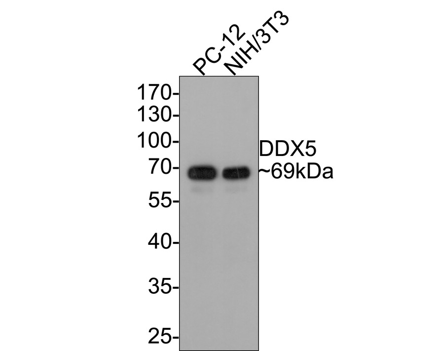
Western blot analysis of DDX5 on different lysates with Rabbit anti-DDX5 antibody at 1/500 dilution. Lane 1: PC-12 cell lysate, Lane 2: NIH/3T3 cell lysate, Lysates/proteins at 10 µg/Lane. Exposure time: 30 seconds; 10% SDS-PAGE gel. Proteins were transferred to a PVDF membrane and blocked with 5% NFDM/TBST for 1 hour at room temperature. The primary antibody at 1/500 dilution was used in 5% NFDM/TBST at room temperature for 2 hours. Goat Anti-Rabbit IgG - HRP Secondary Antibody at 1:300,000 dilution was used for 1 hour at room temperature.WB
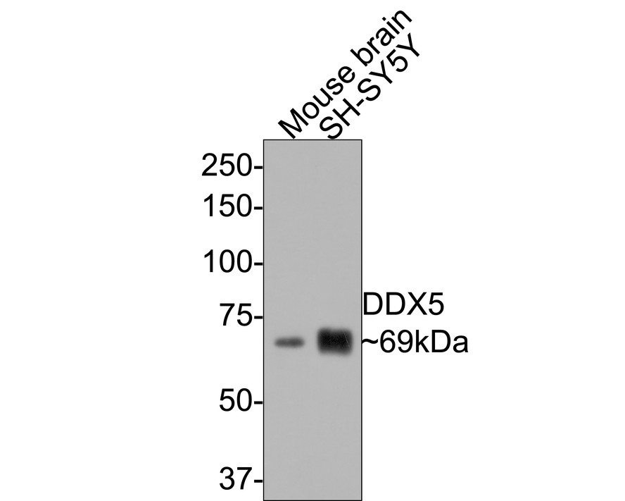
Western blot analysis of DDX5 on different lysates with Rabbit anti-DDX5 antibody at 1/1,000 dilution. Lane 1: Mouse brain tissue lysate, Lane 2: SH-SY5Y cell lysate (10 µg/Lane), Lysates/proteins at 20 µg/Lane. Exposure time: 30 seconds; 8% SDS-PAGE gel. Proteins were transferred to a PVDF membrane and blocked with 5% NFDM/TBST for 1 hour at room temperature. The primary antibody at 1/1,000 dilution was used in 5% NFDM/TBST at room temperature for 2 hours. Goat Anti-Rabbit IgG - HRP Secondary Antibody at 1:300,000 dilution was used for 1 hour at room temperature.IHC
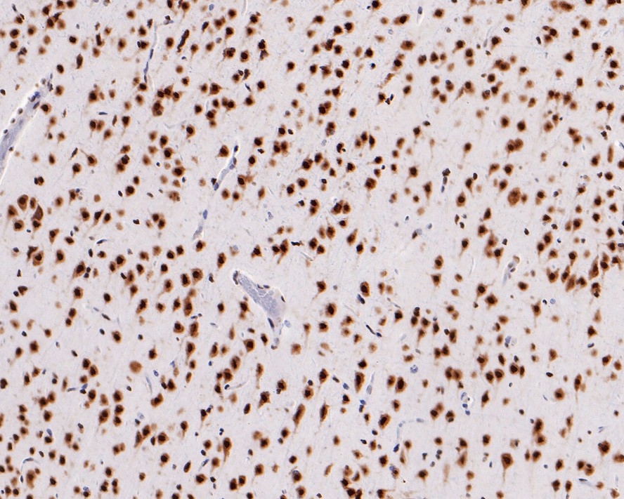
Immunohistochemical analysis of paraffin-embedded rat brain tissue with Rabbit anti-DDX5 antibody at 1/2,000 dilution. The section was pre-treated using heat mediated antigen retrieval with sodium citrate buffer (pH 6.0) for 2 minutes. The tissues were blocked in 1% BSA for 20 minutes at room temperature, washed with ddH2O and PBS, and then probed with the primary antibody at 1/2,000 dilution for 1 hour at room temperature. The detection was performed using an HRP conjugated compact polymer system. DAB was used as the chromogen. Tissues were counterstained with hematoxylin and mounted with DPX.IHC
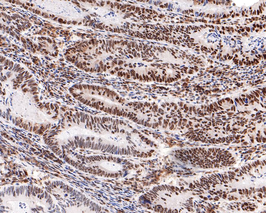
Immunohistochemical analysis of paraffin-embedded human colon carcinoma tissue with Rabbit anti-DDX5 antibody at 1/400 dilution. The section was pre-treated using heat mediated antigen retrieval with sodium citrate buffer (pH 6.0) for 2 minutes. The tissues were blocked in 1% BSA for 20 minutes at room temperature, washed with ddH2O and PBS, and then probed with the primary antibody at 1/400 dilution for 1 hour at room temperature. The detection was performed using an HRP conjugated compact polymer system. DAB was used as the chromogen. Tissues were counterstained with hematoxylin and mounted with DPX.ICC/IF
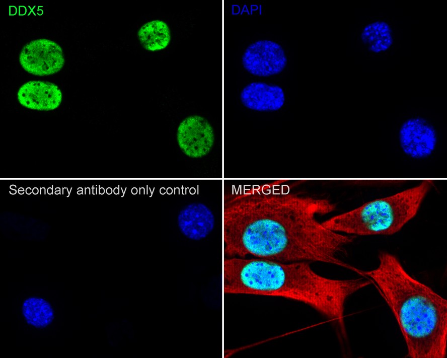
Immunocytochemistry analysis of NIH/3T3 cells labeling DDX5 with Rabbit anti-DDX5 antibody at 1/250 dilution. Cells were fixed in 4% paraformaldehyde for 20 minutes at room temperature, permeabilized with 0.1% Triton X-100 in PBS for 5 minutes at room temperature, then blocked with 1% BSA in 10% negative goat serum for 1 hour at room temperature. Cells were then incubated with Rabbit anti-DDX5 antibody at 1/250 dilution in 1% BSA in PBST overnight at 4 ℃. Goat Anti-Rabbit IgG H&L (488) was used as the secondary antibody at 1/1,000 dilution. PBS instead of the primary antibody was used as the secondary antibody only control. Nuclear DNA was labelled in blue with DAPI. Beta tubulin (red) was stained at 1/100 dilution overnight at +4℃. Goat Anti-Mouse IgG H&L (594) was used as the secondary antibody at 1/1,000 dilution.ICC/IF
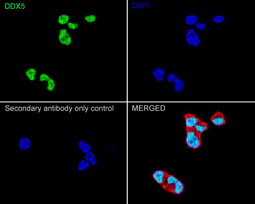
Immunocytochemistry analysis of SH-SY5Y cells labeling DDX5 with Rabbit anti-DDX5 antibody at 1/250 dilution. Cells were fixed in 4% paraformaldehyde for 20 minutes at room temperature, permeabilized with 0.1% Triton X-100 in PBS for 5 minutes at room temperature, then blocked with 1% BSA in 10% negative goat serum for 1 hour at room temperature. Cells were then incubated with Rabbit anti-DDX5 antibody at 1/250 dilution in 1% BSA in PBST overnight at 4 ℃. Goat Anti-Rabbit IgG H&L (488) was used as the secondary antibody at 1/1,000 dilution. PBS instead of the primary antibody was used as the secondary antibody only control. Nuclear DNA was labelled in blue with DAPI. Beta tubulin (red) was stained at 1/100 dilution overnight at +4℃. Goat Anti-Mouse IgG H&L (594) was used as the secondary antibody at 1/1,000 dilution.| Product Name | DDX5 Recombinant Rabbit Monoclonal Antibody |
|---|---|
| Antibody Type | Primary Antibodies |
| Product description | p68 RNA helicase is a nuclear protein that exhibits RNA-dependent ATPase activity. Phosphorylation by protein kinase C inhibits p68 RNA helicase activity. p68 RNA helicase appears to play a role in organ differentiation during development. Furthermore, p68 RNA helicase is expressed in early neural development and in various mesodermal tissues in a number of different chordate embryos. At the cellular level, the expression levels of p68 RNA helicase increases in serum-induced quiescent cell lines. p68 RNA helicase may function as a coactivator for estrogen receptor alpha. Additionally, p68 RNA helicase associates with transcriptional coactivators CBP and p300. p68 RNA helicase localizes to the nucleus under normal conditions. During late telophase, p68 RNA helicase and fibrillarin colocalize to nascent nucleoli. p68 RNA helicase may function as a heterodimer with p72 RNA helicase. |
| Immunogen | Synthetic peptide within Human DDX5 aa 1-50 / 614. |
| Clonality | monoclonal |
|---|---|
| Isotype | IgG |
| Host Species | Recombinant rabbit |
| Tested Applications | ICC/IFIHCWB |
| WB:1:500-1:2000 IHC:1:50-1:2000 ICC/IF:1:50-1:200 |
|
| Species Reactivity | HumanMouseRat |
| Concentration | 1mg/ml |
| Purification | Protein A |
| Alternative Names | ATP dependent RNA helicase DDX5 antibody
DDX 5 antibody
Ddx5 antibody
DDX5_HUMAN antibody
DEAD (Asp Glu Ala Asp) box helicase 5 antibody
DEAD (Asp Glu Ala Asp) box polypeptide 5 antibody
DEAD box 5 antibody
DEAD box protein 5 antibody
DEAD/H (Asp Glu Ala Asp/His) box polypeptide 5 (RNA helicase 68kD) antibody G17P1 antibody HELR antibody HLR1 antibody HUMP68 antibody P68 antibody p68 RNA helicase antibody Probable ATP dependent RNA helicase DDX5 antibody Probable ATP-dependent RNA helicase DDX5 antibody RNA helicase p68 antibody |
|---|---|
| Molecular Weight(MW) | 69kDa |
| Cellular Localization | Nucleus. |
WB
Western blot analysis of DDX5 on different lysates with Rabbit anti-DDX5 antibody at 1/500 dilution. Lane 1: PC-12 cell lysate, Lane 2: NIH/3T3 cell lysate, Lysates/proteins at 10 µg/Lane. Exposure time: 30 seconds; 10% SDS-PAGE gel. Proteins were transferred to a PVDF membrane and blocked with 5% NFDM/TBST for 1 hour at room temperature. The primary antibody at 1/500 dilution was used in 5% NFDM/TBST at room temperature for 2 hours. Goat Anti-Rabbit IgG - HRP Secondary Antibody at 1:300,000 dilution was used for 1 hour at room temperature.
WB
Western blot analysis of DDX5 on different lysates with Rabbit anti-DDX5 antibody at 1/1,000 dilution. Lane 1: Mouse brain tissue lysate, Lane 2: SH-SY5Y cell lysate (10 µg/Lane), Lysates/proteins at 20 µg/Lane. Exposure time: 30 seconds; 8% SDS-PAGE gel. Proteins were transferred to a PVDF membrane and blocked with 5% NFDM/TBST for 1 hour at room temperature. The primary antibody at 1/1,000 dilution was used in 5% NFDM/TBST at room temperature for 2 hours. Goat Anti-Rabbit IgG - HRP Secondary Antibody at 1:300,000 dilution was used for 1 hour at room temperature.
IHC
Immunohistochemical analysis of paraffin-embedded rat brain tissue with Rabbit anti-DDX5 antibody at 1/2,000 dilution. The section was pre-treated using heat mediated antigen retrieval with sodium citrate buffer (pH 6.0) for 2 minutes. The tissues were blocked in 1% BSA for 20 minutes at room temperature, washed with ddH2O and PBS, and then probed with the primary antibody at 1/2,000 dilution for 1 hour at room temperature. The detection was performed using an HRP conjugated compact polymer system. DAB was used as the chromogen. Tissues were counterstained with hematoxylin and mounted with DPX.
IHC
Immunohistochemical analysis of paraffin-embedded human colon carcinoma tissue with Rabbit anti-DDX5 antibody at 1/400 dilution. The section was pre-treated using heat mediated antigen retrieval with sodium citrate buffer (pH 6.0) for 2 minutes. The tissues were blocked in 1% BSA for 20 minutes at room temperature, washed with ddH2O and PBS, and then probed with the primary antibody at 1/400 dilution for 1 hour at room temperature. The detection was performed using an HRP conjugated compact polymer system. DAB was used as the chromogen. Tissues were counterstained with hematoxylin and mounted with DPX.
ICC/IF
Immunocytochemistry analysis of NIH/3T3 cells labeling DDX5 with Rabbit anti-DDX5 antibody at 1/250 dilution. Cells were fixed in 4% paraformaldehyde for 20 minutes at room temperature, permeabilized with 0.1% Triton X-100 in PBS for 5 minutes at room temperature, then blocked with 1% BSA in 10% negative goat serum for 1 hour at room temperature. Cells were then incubated with Rabbit anti-DDX5 antibody at 1/250 dilution in 1% BSA in PBST overnight at 4 ℃. Goat Anti-Rabbit IgG H&L (488) was used as the secondary antibody at 1/1,000 dilution. PBS instead of the primary antibody was used as the secondary antibody only control. Nuclear DNA was labelled in blue with DAPI. Beta tubulin (red) was stained at 1/100 dilution overnight at +4℃. Goat Anti-Mouse IgG H&L (594) was used as the secondary antibody at 1/1,000 dilution.
ICC/IF
Immunocytochemistry analysis of SH-SY5Y cells labeling DDX5 with Rabbit anti-DDX5 antibody at 1/250 dilution. Cells were fixed in 4% paraformaldehyde for 20 minutes at room temperature, permeabilized with 0.1% Triton X-100 in PBS for 5 minutes at room temperature, then blocked with 1% BSA in 10% negative goat serum for 1 hour at room temperature. Cells were then incubated with Rabbit anti-DDX5 antibody at 1/250 dilution in 1% BSA in PBST overnight at 4 ℃. Goat Anti-Rabbit IgG H&L (488) was used as the secondary antibody at 1/1,000 dilution. PBS instead of the primary antibody was used as the secondary antibody only control. Nuclear DNA was labelled in blue with DAPI. Beta tubulin (red) was stained at 1/100 dilution overnight at +4℃. Goat Anti-Mouse IgG H&L (594) was used as the secondary antibody at 1/1,000 dilution.| Application Notes | WB:1:500-1:2000 IHC:1:50-1:2000 ICC/IF:1:50-1:200 |
|---|
| Form | Liquid |
|---|---|
| Storage Instructions | Store at +4℃ after thawing. Aliquot store at -20℃ or -80℃. Avoid repeated freeze / thaw cycles. |
| Storage Buffer | 1*TBS (pH7.4), 1%BSA, 40%Glycerol. Preservative: 0.05% Sodium Azide. |


