Application
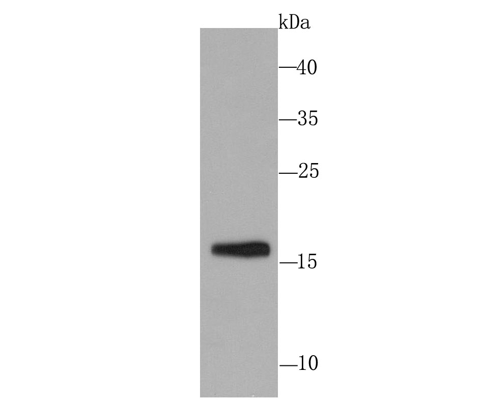
Fig1: Western blot analysis of Skp1 on PC-12 cell using anti-Skp1 antibody at 1/1,000 dilution.Application
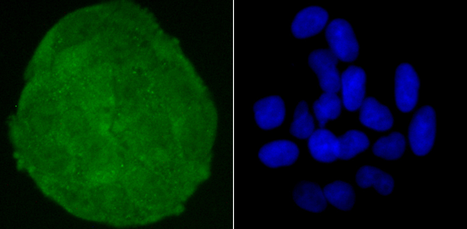
Fig2: ICC staining Skp1 in Hela cells (green). The nuclear counter stain is DAPI (blue). Cells were fixed in paraformaldehyde, permeabilised with 0.25% Triton X100/PBS.Application
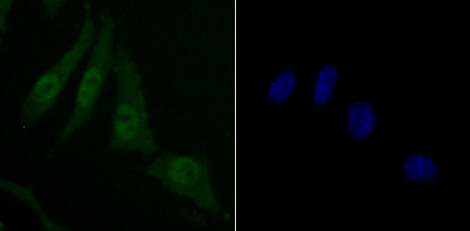
Fig3: ICC staining Skp1 in SH-SY5Y cells (green). The nuclear counter stain is DAPI (blue). Cells were fixed in paraformaldehyde, permeabilised with 0.25% Triton X100/PBS.Application
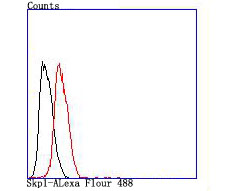
Fig4: Flow cytometric analysis of Hela cells with Skp1 antibody at 1/100 dilution (red) compared with an unlabelled control (cells without incubation with primary antibody; black).| Product Name | Skp1 [JM27-37] |
|---|---|
| Antibody Type | Primary Antibodies |
| Antigen Alias | Cyclin A/CDK2 associated p19 antibody Cyclin A/CDK2 associated protein p19 antibody Cyclin-A/CDK2-associated protein p19 antibody EMC19 antibody MGC34403 antibody OCP 2 antibody OCP II antibody OCP II protein antibody OCP-2 antibody OCP-II antibody OCP2 antibody OCPII antibody Organ of Corti protein 2 antibody Organ of Corti protein II antibody OTTHUMP00000159379 antibody OTTHUMP00000159380 antibody OTTHUMP00000228275 antibody OTTHUMP00000228276 antibody OTTHUMP00000228278 antibody OTTHUMP00000228279 antibody OTTHUMP00000228281 antibody p19A antibody p19skp1 antibody RNA polymerase II elongation factor like protein antibody RNA polymerase II elongation factor like protein OCP2 antibody RNA polymerase II elongation factor-like protein antibody S phase kinase associated protein 1 antibody S phase kinase associated protein 1A antibody S phase kinase associated protein 1A p19A antibody S-phase kinase-associated protein 1 antibody SIII antibody Skp 1 antibody SKP 1A antibody skp1 antibody SKP1_HUMAN antibody SKP1A antibody TCEB1L antibody Transcription Elongation Factor B 1 like antibody Transcription elongation factor B antibody Transcription elongation factor B SIII polypeptide 1 like antibody |
| Product description | Essential component of the SCF (SKP1-CUL1-F-box protein) ubiquitin ligase complex, which mediates the ubiquitination of proteins involved in cell cycle progression, signal transduction and transcription. In the SCF complex, serves as an adapter that links the F-box protein to CUL1. SCF(BTRC) mediates the ubiquitination of NFKBIA at 'Lys-21' and 'Lys-22'; the degradation frees the associated NFKB1-RELA dimer to translocate into the nucleus and to activate transcription. SCF(Cyclin F) directs ubiquitination of CP110. |
| Immunogen | recombinant protein |
| Clonality | Monoclonal |
|---|---|
| Isotype | IgG |
| Host Species | Recombinant rabbit |
| Tested Applications | WBICC/IFFC |
| WB:1:500 ICC:1:50-1:100 FC:1:50-1:100 |
|
| Species Reactivity | HumanMouseRat |
| Concentration | 1mg/ml |
| Alternative Names | Cyclin A/CDK2 associated p19 antibody Cyclin A/CDK2 associated protein p19 antibody Cyclin-A/CDK2-associated protein p19 antibody EMC19 antibody MGC34403 antibody OCP 2 antibody OCP II antibody OCP II protein antibody OCP-2 antibody OCP-II antibody OCP2 antibody OCPII antibody Organ of Corti protein 2 antibody Organ of Corti protein II antibody OTTHUMP00000159379 antibody OTTHUMP00000159380 antibody OTTHUMP00000228275 antibody OTTHUMP00000228276 antibody OTTHUMP00000228278 antibody OTTHUMP00000228279 antibody OTTHUMP00000228281 antibody p19A antibody p19skp1 antibody RNA polymerase II elongation factor like protein antibody RNA polymerase II elongation factor like protein OCP2 antibody RNA polymerase II elongation factor-like protein antibody S phase kinase associated protein 1 antibody S phase kinase associated protein 1A antibody S phase kinase associated protein 1A p19A antibody S-phase kinase-associated protein 1 antibody SIII antibody Skp 1 antibody SKP 1A antibody skp1 antibody SKP1_HUMAN antibody SKP1A antibody TCEB1L antibody Transcription Elongation Factor B 1 like antibody Transcription elongation factor B antibody Transcription elongation factor B SIII polypeptide 1 like antibody |
|---|---|
| Molecular Weight(MW) | 19 kDa |
| Cellular Localization | Cytoplasm. Nucleus. |
Application
Fig1: Western blot analysis of Skp1 on PC-12 cell using anti-Skp1 antibody at 1/1,000 dilution.
Application
Fig2: ICC staining Skp1 in Hela cells (green). The nuclear counter stain is DAPI (blue). Cells were fixed in paraformaldehyde, permeabilised with 0.25% Triton X100/PBS.
Application
Fig3: ICC staining Skp1 in SH-SY5Y cells (green). The nuclear counter stain is DAPI (blue). Cells were fixed in paraformaldehyde, permeabilised with 0.25% Triton X100/PBS.
Application
Fig4: Flow cytometric analysis of Hela cells with Skp1 antibody at 1/100 dilution (red) compared with an unlabelled control (cells without incubation with primary antibody; black).| Positive Control | PC-12, Hela, SH-SY5Y. |
|---|---|
| Application Notes | WB:1:500 ICC:1:50-1:100 FC:1:50-1:100 |
| Form | Liquid |
|---|---|
| Storage Instructions | Store at +4℃ after thawing. Aliquot store at -20℃ or -80℃. Avoid repeated freeze / thaw cycles. |
| Storage Buffer | 1*TBS (pH7.4), 1%BSA, 40%Glycerol. Preservative: 0.05% Sodium Azide. |


