WB
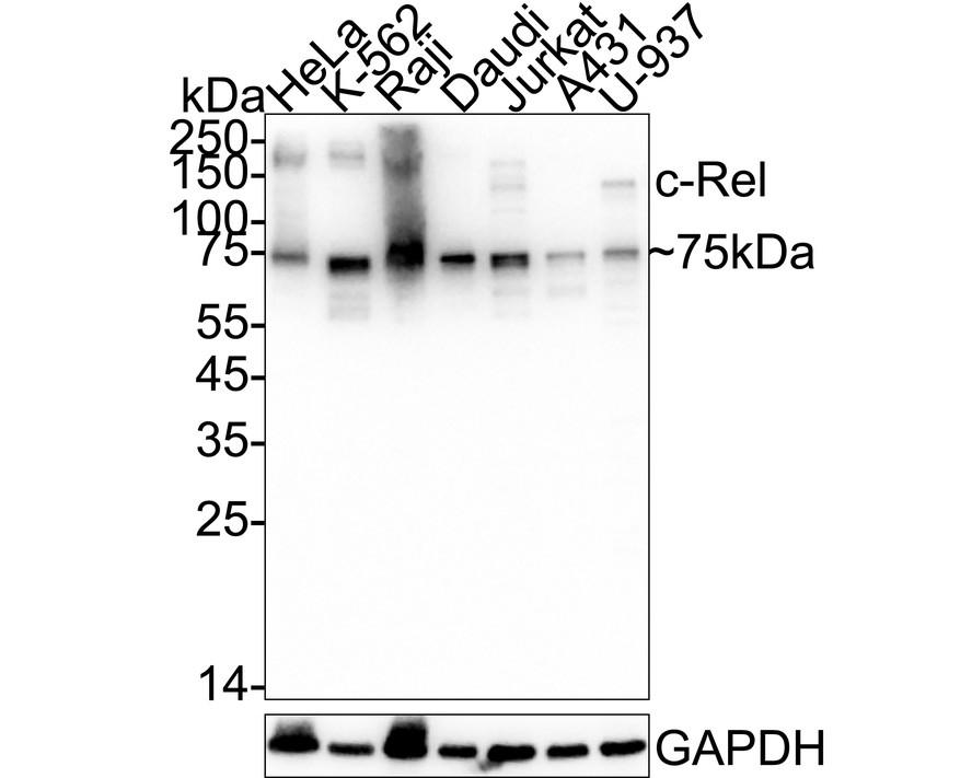
Western blot analysis of c-Rel on different lysates with Rabbit anti-c-Rel antibody at 1/1,000 dilution. Lane 1: HeLa cell lysate, Lane 2: K-562 cell lysate, Lane 3: Raji cell lysate, Lane 4: Daudi cell lysate, Lane 5: Jurkat cell lysate, Lane 6: A431 cell lysate, Lane 7: U-937 cell lysate, Lysates/proteins at 20 µg/Lane. Exposure time: 25 seconds; 4-20% SDS-PAGE gel. Proteins were transferred to a PVDF membrane and blocked with 5% NFDM/TBST for 1 hour at room temperature. The primary antibody at 1/1,000 dilution was used in 5% NFDM/TBST at 4℃ overnight. Goat Anti-Rabbit IgG - HRP Secondary Antibody at 1/50,000 dilution was used for 1 hour at room temperature.IHC
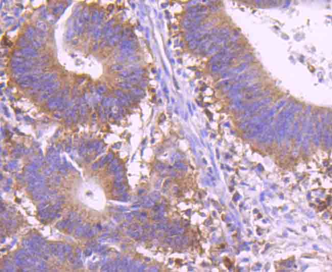
Immunohistochemical analysis of paraffin-embedded human colon cancer tissue using anti-c-Rel antibody. Counter stained with hematoxylin.IHC
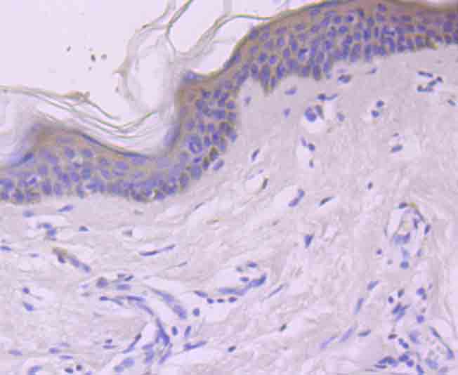
Immunohistochemical analysis of paraffin-embedded human skin tissue using anti-c-Rel antibody. Counter stained with hematoxylin.IHC
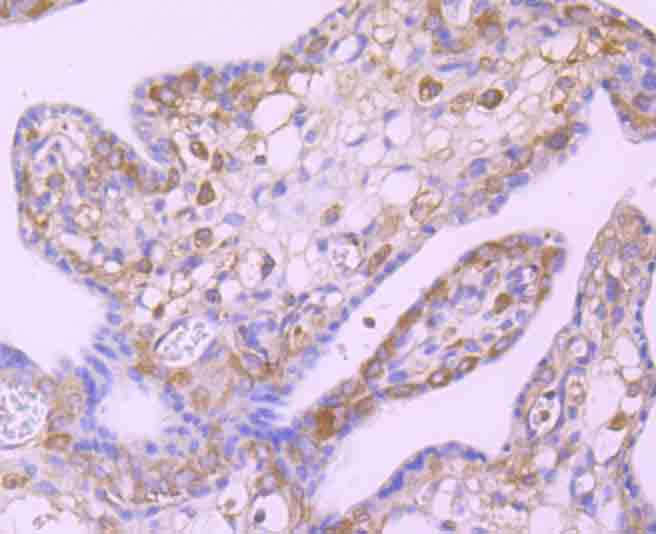
Immunohistochemical analysis of paraffin-embedded human placenta tissue using anti-c-Rel antibody. Counter stained with hematoxylin.| Product Name | c-Rel Recombinant Rabbit Monoclonal Antibody |
|---|---|
| Antibody Type | Primary Antibodies |
| Product description | c-Rel is the cellular cognate of v-Rel, the avian reticuloendotheliosis virus strain T transforming gene. v-Rel encodes a phosphoprotein that is located in the cytoplasm of transformed spleen cells and in the nucleus of non-transformed fibroblasts, in contrast to the c-Rel protein, which is cytoplasmic. c-Rel has been shown to represent a constituent of the κB site binding transcription factor NFκB, which plays a crucial role in the expression of immunoglobulin κ light chain gene. In contrast to c-Rel, v-Rel is truncated in its C-terminal transactivation domain and does not appear to function as a transcriptional transactivator. It has thus been postulated that v-Rel may interfere with the normal transcription of NFκB regulated genes and thus cause transformation by a mechanism analogous to v-ErbA, which binds to the thyroid hormone-responsive region in certain erythroid genes needed for differentiation, but cannot be activated by thyroid hormone. |
| Immunogen | Synthetic peptide within Human c-Rel aa 501-550 / 619. |
| Clonality | Monoclonal |
|---|---|
| Isotype | IgG |
| Host Species | Recombinant rabbit |
| Tested Applications | IHCWB |
| WB:1:1000 IHC:1:50-1:200 |
|
| Species Reactivity | Human |
| Concentration | 1mg/ml |
| Purification | Protein A |
| Alternative Names | Avian reticuloendotheliosis antibody C REL antibody C Rel protein antibody c Rel proto oncogene protein antibody Oncogene REL antibody Oncogene REL avian reticuloendotheliosis antibody Proto-oncogene c-Rel antibody REL antibody REL_HUMAN antibody v rel avian reticuloendotheliosis viral oncogene homolog antibody v rel reticuloendotheliosis viral oncogene homolog antibody V rel reticuloendotheliosis viral oncogene homolog (avian) antibody |
|---|---|
| Molecular Weight(MW) | 69kDa(Observed band size: 75kDa) |
| Cellular Localization | Cytoplasm. Nucleus. |
| SwissProt ID | Q04864 |
|---|
WB
Western blot analysis of c-Rel on different lysates with Rabbit anti-c-Rel antibody at 1/1,000 dilution. Lane 1: HeLa cell lysate, Lane 2: K-562 cell lysate, Lane 3: Raji cell lysate, Lane 4: Daudi cell lysate, Lane 5: Jurkat cell lysate, Lane 6: A431 cell lysate, Lane 7: U-937 cell lysate, Lysates/proteins at 20 µg/Lane. Exposure time: 25 seconds; 4-20% SDS-PAGE gel. Proteins were transferred to a PVDF membrane and blocked with 5% NFDM/TBST for 1 hour at room temperature. The primary antibody at 1/1,000 dilution was used in 5% NFDM/TBST at 4℃ overnight. Goat Anti-Rabbit IgG - HRP Secondary Antibody at 1/50,000 dilution was used for 1 hour at room temperature.
IHC
Immunohistochemical analysis of paraffin-embedded human colon cancer tissue using anti-c-Rel antibody. Counter stained with hematoxylin.
IHC
Immunohistochemical analysis of paraffin-embedded human skin tissue using anti-c-Rel antibody. Counter stained with hematoxylin.
IHC
Immunohistochemical analysis of paraffin-embedded human placenta tissue using anti-c-Rel antibody. Counter stained with hematoxylin.| Application Notes | WB:1:1000 IHC:1:50-1:200 |
|---|
| Form | Liquid |
|---|---|
| Storage Instructions | Store at +4℃ after thawing. Aliquot store at -20℃ or -80℃. Avoid repeated freeze / thaw cycles. |
| Storage Buffer | 1*TBS (pH7.4), 1%BSA, 40%Glycerol. Preservative: 0.05% Sodium Azide. |


