Application
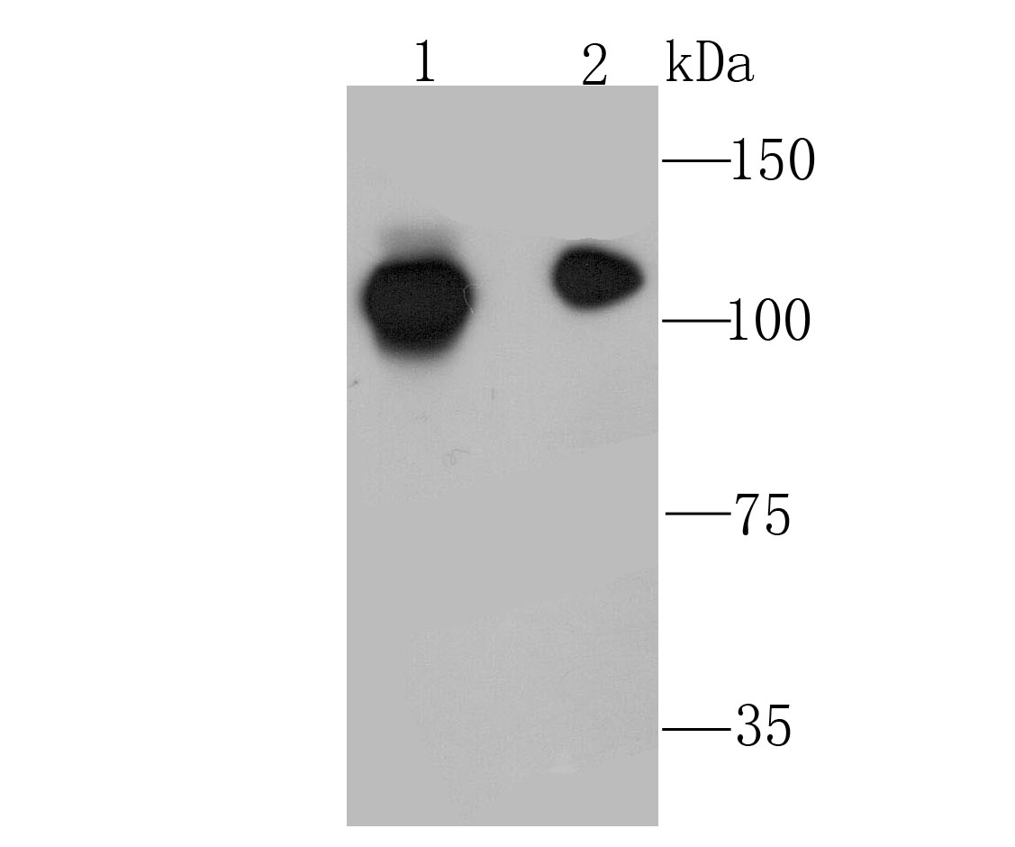
Fig1: Western blot analysis of NFkB p100 on Hela (1) and MCF-7 (2) cell using anti-NFkB p100 antibody at 1/1,000 dilution.Application
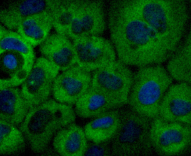
Fig2: ICC staining NFkB p100 in A431 cells (green). The nuclear counter stain is DAPI (blue). Cells were fixed in paraformaldehyde, permeabilised with 0.25% Triton X100/PBS.Application
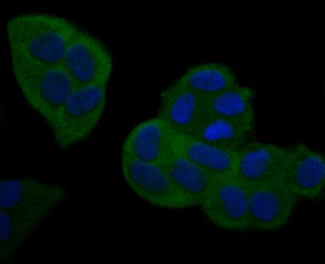
Fig3: ICC staining NFkB p100 in Hela cells (green). The nuclear counter stain is DAPI (blue). Cells were fixed in paraformaldehyde, permeabilised with 0.25% Triton X100/PBS.Application
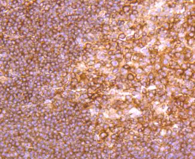
Fig4: Immunohistochemical analysis of paraffin-embedded human tonsil tissue using anti-NFkB p100 antibody. Counter stained with hematoxylin.Application
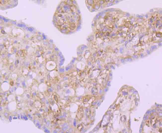
Fig5: Immunohistochemical analysis of paraffin-embedded human placenta tissue using anti-NFkB p100 antibody. Counter stained with hematoxylin.Application
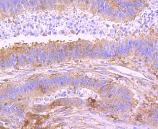
Fig6: Immunohistochemical analysis of paraffin-embedded human colon cancer tissue using anti-NFkB p100 antibody. Counter stained with hematoxylin.| Product Name | NFkB p100 [JM82-03] |
|---|---|
| Antibody Type | Primary Antibodies |
| Antigen Alias | CVID10 antibody DNA binding factor KBF2 antibody H2TF1 antibody Lymphocyte translocation chromosome 10 protein antibody LYT 10 antibody NF kB2 antibody NFKB p52/p100 subunit antibody Nuclear factor Kappa B subunit 2 antibody Nuclear factor of kappa light polypeptide gene enhancer in B cells 2 (p49/p100) antibody Nuclear factor of kappa light polypeptide gene enhancer in B cells 2 antibody Oncogene Lyt 10 antibody p100 antibody Transcription factor NFKB2 antibody |
| Product description | NFKB2 appears to have dual functions such as cytoplasmic retention of attached NF-kappa-B proteins by p100 and generation of p52 by a cotranslational processing. The proteasome-mediated process ensures the production of both p52 and p100 and preserves their independent function. p52 binds to the kappa-B consensus sequence 5'-GGRNNYYCC-3', located in the enhancer region of genes involved in immune response and acute phase reactions. p52 and p100 are respectively the minor and major form; the processing of p100 being relatively poor. Isoform p49 is a subunit of the NF-kappa-B protein complex, which stimulates the HIV enhancer in synergy with p65. |
| Immunogen | Recombinant protein |
| Clonality | Monoclonal |
|---|---|
| Isotype | IgG |
| Host Species | Recombinant rabbit |
| Tested Applications | WBICC/IFIHCIP |
| WB:1:500-1:2,000 ICC:1:50-1:200 IHC:1:50-1:200 |
|
| Species Reactivity | HumanMouseRat |
| Concentration | 1mg/ml |
| Alternative Names | CVID10 antibody DNA binding factor KBF2 antibody H2TF1 antibody Lymphocyte translocation chromosome 10 protein antibody LYT 10 antibody NF kB2 antibody NFKB p52/p100 subunit antibody Nuclear factor Kappa B subunit 2 antibody Nuclear factor of kappa light polypeptide gene enhancer in B cells 2 (p49/p100) antibody Nuclear factor of kappa light polypeptide gene enhancer in B cells 2 antibody Oncogene Lyt 10 antibody p100 antibody Transcription factor NFKB2 antibody |
|---|---|
| Molecular Weight(MW) | 100 kDa |
| Cellular Localization | Cytoplasm. Nucleus. |
Application
Fig1: Western blot analysis of NFkB p100 on Hela (1) and MCF-7 (2) cell using anti-NFkB p100 antibody at 1/1,000 dilution.
Application
Fig2: ICC staining NFkB p100 in A431 cells (green). The nuclear counter stain is DAPI (blue). Cells were fixed in paraformaldehyde, permeabilised with 0.25% Triton X100/PBS.
Application
Fig3: ICC staining NFkB p100 in Hela cells (green). The nuclear counter stain is DAPI (blue). Cells were fixed in paraformaldehyde, permeabilised with 0.25% Triton X100/PBS.
Application
Fig4: Immunohistochemical analysis of paraffin-embedded human tonsil tissue using anti-NFkB p100 antibody. Counter stained with hematoxylin.
Application
Fig5: Immunohistochemical analysis of paraffin-embedded human placenta tissue using anti-NFkB p100 antibody. Counter stained with hematoxylin.
Application
Fig6: Immunohistochemical analysis of paraffin-embedded human colon cancer tissue using anti-NFkB p100 antibody. Counter stained with hematoxylin.| Positive Control | Hela, MCF-7, A431, human tonsil tissue, human placenta tissue, human colon cancer tissue. |
|---|---|
| Application Notes | WB:1:500-1:2,000 ICC:1:50-1:200 IHC:1:50-1:200 |
| Form | Liquid |
|---|---|
| Storage Instructions | Store at +4℃ after thawing. Aliquot store at -20℃ or -80℃. Avoid repeated freeze / thaw cycles. |
| Storage Buffer | 1*TBS (pH7.4), 1%BSA, 40%Glycerol. Preservative: 0.05% Sodium Azide. |


