WB
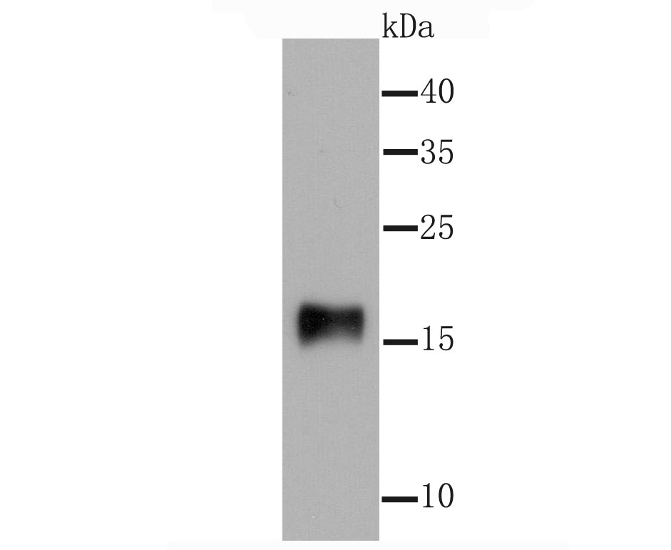
Western blot analysis of gamma Synuclein on rat kidney tissue lysate using anti-gamma Synuclein antibody at 1/1,000 dilution.ICC/IF
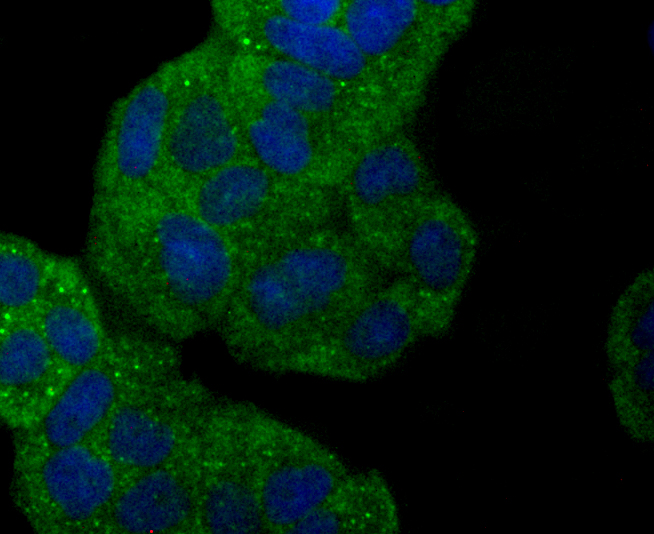
ICC staining gamma Synuclein in Hela cells (green). The nuclear counter stain is DAPI (blue). Cells were fixed in paraformaldehyde, permeabilised with 0.25% Triton X100/PBS.IHC
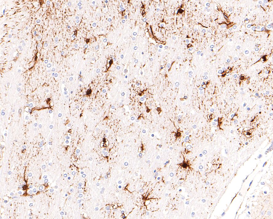
Immunohistochemical analysis of paraffin-embedded rat brain tissue with Rabbit anti-gamma Synuclein antibody at 1/400 dilution. The section was pre-treated using heat mediated antigen retrieval with Tris-EDTA buffer (pH 9.0) for 20 minutes. The tissues were blocked in 1% BSA for 20 minutes at room temperature, washed with ddH2O and PBS, and then probed with the primary antibody at 1/400 dilution for 1 hour at room temperature. The detection was performed using an HRP conjugated compact polymer system. DAB was used as the chromogen. Tissues were counterstained with hematoxylin and mounted with DPX.IHC
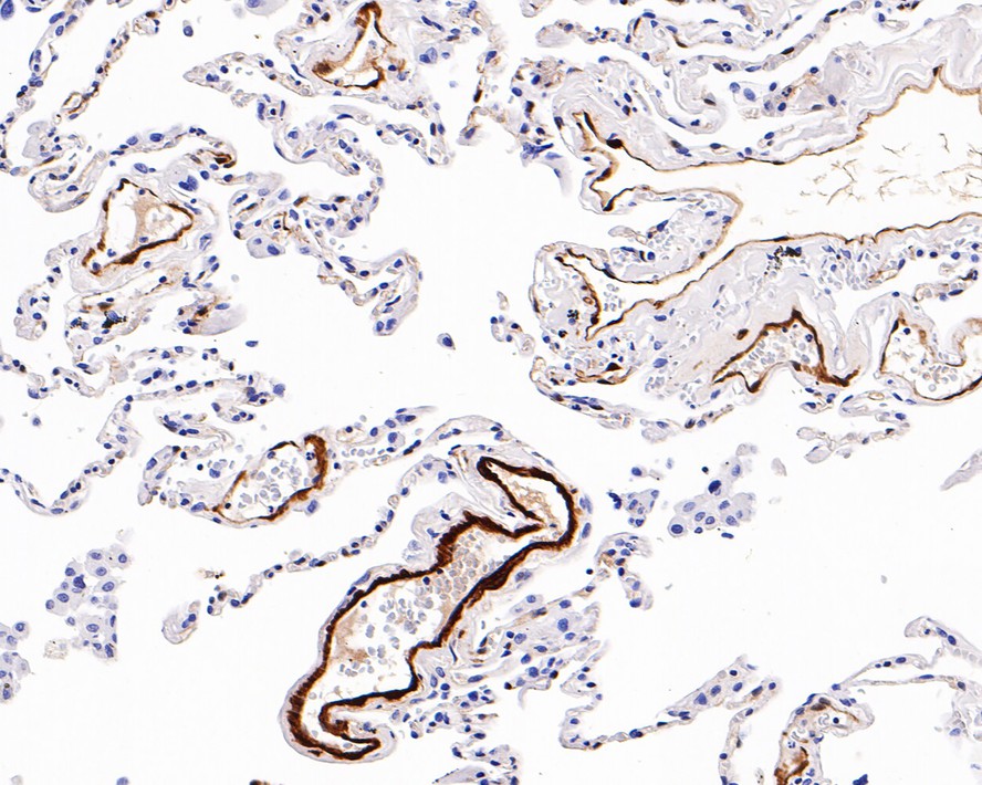
Immunohistochemical analysis of paraffin-embedded human lung tissue with Rabbit anti-gamma Synuclein antibody at 1/1,000 dilution. The section was pre-treated using heat mediated antigen retrieval with Tris-EDTA buffer (pH 9.0) for 20 minutes. The tissues were blocked in 1% BSA for 20 minutes at room temperature, washed with ddH2O and PBS, and then probed with the primary antibody at 1/1,000 dilution for 1 hour at room temperature. The detection was performed using an HRP conjugated compact polymer system. DAB was used as the chromogen. Tissues were counterstained with hematoxylin and mounted with DPX.IHC
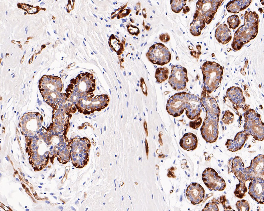
Immunohistochemical analysis of paraffin-embedded human breast tissue with Rabbit anti-gamma Synuclein antibody at 1/1,000 dilution. The section was pre-treated using heat mediated antigen retrieval with Tris-EDTA buffer (pH 9.0) for 20 minutes. The tissues were blocked in 1% BSA for 20 minutes at room temperature, washed with ddH2O and PBS, and then probed with the primary antibody at 1/1,000 dilution for 1 hour at room temperature. The detection was performed using an HRP conjugated compact polymer system. DAB was used as the chromogen. Tissues were counterstained with hematoxylin and mounted with DPX.FC
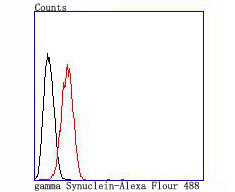
Flow cytometric analysis of Hela cells with gamma Synuclein antibody at 1/100 dilution (red) compared with an unlabelled control (cells without incubation with primary antibody; black).| Product Name | gamma Synuclein Recombinant Rabbit Monoclonal Antibody |
|---|---|
| Antibody Type | Primary Antibodies |
| Product description | The synucleins, including α-synuclein (also designated NACP for nonamyloid component precursor), β-synuclein (also designated PNP 14 for phospho-neuroprotein 14) and γ-synuclein (also designated persyn or BCSG1 for breast cancer-specific gene 1) are presynaptic proteins abundant in neurons. Synucleins are predominantly expressed in the brain and are speculated to be involved in synaptic regulation and neuronal plasticity. α-Synuclein, identified as a component of Alzheimer's disease amyloid plaques, is localized to neuronal cell bodies and synapses. Coordinate expression of α-synuclein and β-synuclein may be important during hematopoetic cell differentiation. A mutant form of α-synuclein is found in patients with early onset Parkinson's disease. γ-Synuclein is associated with axonal pathology in Parkinson's disease. |
| Immunogen | Synthetic peptide within Human gamma Synuclein aa 78-127 / 127. |
| Clonality | Monoclonal |
|---|---|
| Isotype | IgG |
| Host Species | Recombinant rabbit |
| Tested Applications | FCICC/IFIHCWB |
| WB:1:500-1:2000 IHC:1:400-1:1000 ICC/IF:1:50-1:200 FC:1:50-1:100 |
|
| Species Reactivity | HumanRat |
| Concentration | 1mg/ml |
| Purification | Protein A |
| Alternative Names | BCSG1 antibody
Breast cancer specific gene 1 protein antibody
Breast cancer specific protein 1 antibody
Breast cancer-specific gene 1 protein antibody
Gamma synuclein antibody
Gamma-synuclein antibody
Persyn antibody
PRSN antibody
SNCG antibody
SR antibody
Synoretin antibody
Synuclein gamma antibody
synuclein gamma (breast cancer-specific protein 1) antibody SYUG_HUMAN antibody |
|---|---|
| Molecular Weight(MW) | 16 kDa |
| Cellular Localization | Cytoplasm. Cytoskeleton. Centrosome. |
WB
Western blot analysis of gamma Synuclein on rat kidney tissue lysate using anti-gamma Synuclein antibody at 1/1,000 dilution.
ICC/IF
ICC staining gamma Synuclein in Hela cells (green). The nuclear counter stain is DAPI (blue). Cells were fixed in paraformaldehyde, permeabilised with 0.25% Triton X100/PBS.
IHC
Immunohistochemical analysis of paraffin-embedded rat brain tissue with Rabbit anti-gamma Synuclein antibody at 1/400 dilution. The section was pre-treated using heat mediated antigen retrieval with Tris-EDTA buffer (pH 9.0) for 20 minutes. The tissues were blocked in 1% BSA for 20 minutes at room temperature, washed with ddH2O and PBS, and then probed with the primary antibody at 1/400 dilution for 1 hour at room temperature. The detection was performed using an HRP conjugated compact polymer system. DAB was used as the chromogen. Tissues were counterstained with hematoxylin and mounted with DPX.
IHC
Immunohistochemical analysis of paraffin-embedded human lung tissue with Rabbit anti-gamma Synuclein antibody at 1/1,000 dilution. The section was pre-treated using heat mediated antigen retrieval with Tris-EDTA buffer (pH 9.0) for 20 minutes. The tissues were blocked in 1% BSA for 20 minutes at room temperature, washed with ddH2O and PBS, and then probed with the primary antibody at 1/1,000 dilution for 1 hour at room temperature. The detection was performed using an HRP conjugated compact polymer system. DAB was used as the chromogen. Tissues were counterstained with hematoxylin and mounted with DPX.
IHC
Immunohistochemical analysis of paraffin-embedded human breast tissue with Rabbit anti-gamma Synuclein antibody at 1/1,000 dilution. The section was pre-treated using heat mediated antigen retrieval with Tris-EDTA buffer (pH 9.0) for 20 minutes. The tissues were blocked in 1% BSA for 20 minutes at room temperature, washed with ddH2O and PBS, and then probed with the primary antibody at 1/1,000 dilution for 1 hour at room temperature. The detection was performed using an HRP conjugated compact polymer system. DAB was used as the chromogen. Tissues were counterstained with hematoxylin and mounted with DPX.
FC
Flow cytometric analysis of Hela cells with gamma Synuclein antibody at 1/100 dilution (red) compared with an unlabelled control (cells without incubation with primary antibody; black).| Application Notes | WB:1:500-1:2000 IHC:1:400-1:1000 ICC/IF:1:50-1:200 FC:1:50-1:100 |
|---|
| Form | Liquid |
|---|---|
| Storage Instructions | Store at +4℃ after thawing. Aliquot store at -20℃ or -80℃. Avoid repeated freeze / thaw cycles. |
| Storage Buffer | 1*TBS (pH7.4), 1%BSA, 40%Glycerol. Preservative: 0.05% Sodium Azide. |


