Application
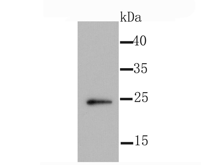
Fig1: Western blot analysis of Id1 on 293T cell using anti-Id1 antibody at 1/1,000 dilution.Application
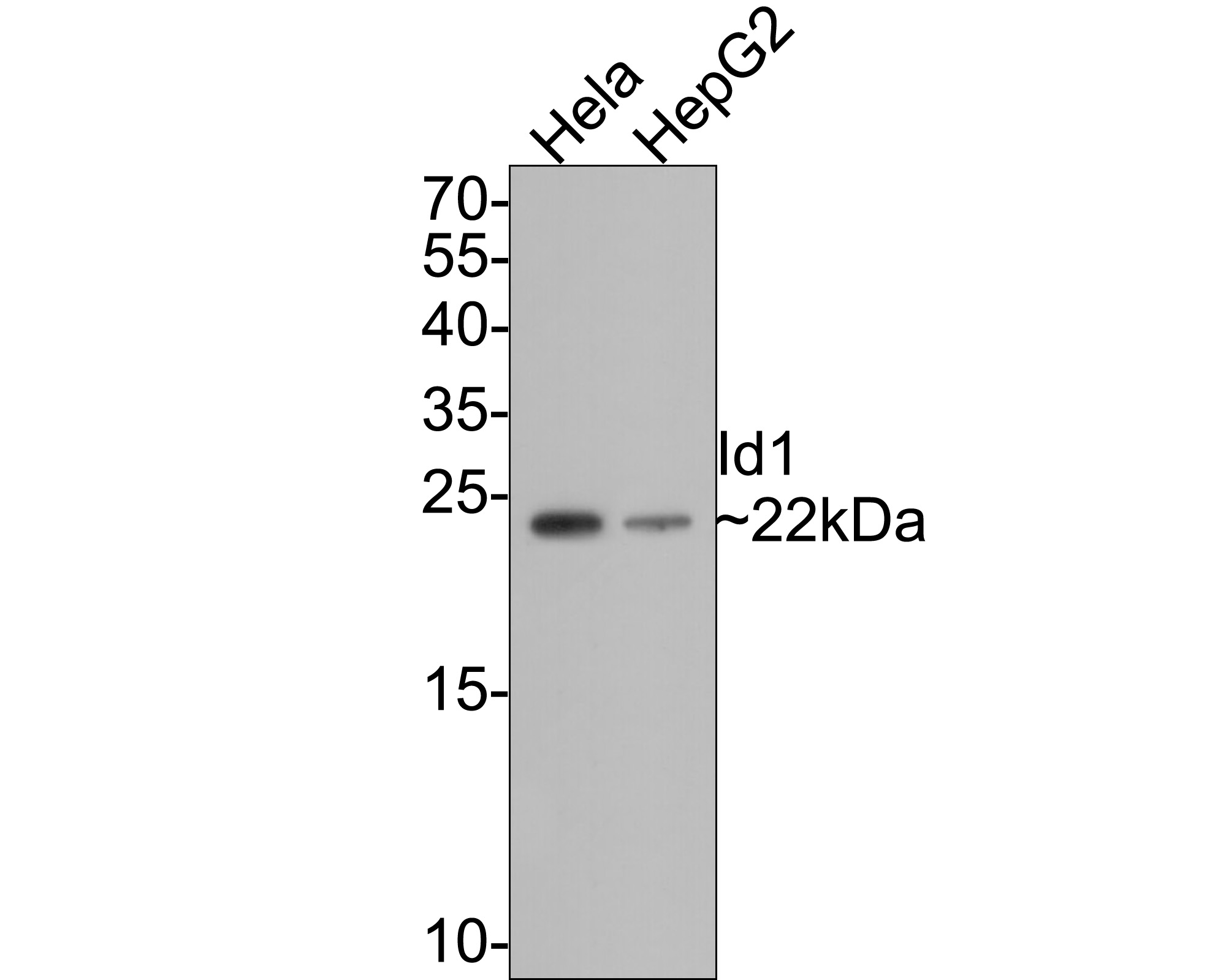
Fig2: Western blot analysis of Id1 on different lysates with Rabbit anti-Id1 antibody at 1/500 dilution. Lane 1: Hela cell lysate, Lane 2: HepG2 cell lysate, Lysates/proteins at 10 µg/Lane. Exposure time: 1 minute; 15% SDS-PAGE gel. Proteins were transferred to a PVDF membrane and blocked with 5% NFDM/TBST for 1 hour at room temperature. The primary antibody at 1/500 dilution was used in 5% NFDM/TBST at room temperature for 2 hours. Goat Anti-Rabbit IgG - HRP Secondary Antibody at 1:300,000 dilution was used for 1 hour at room temperature.Application
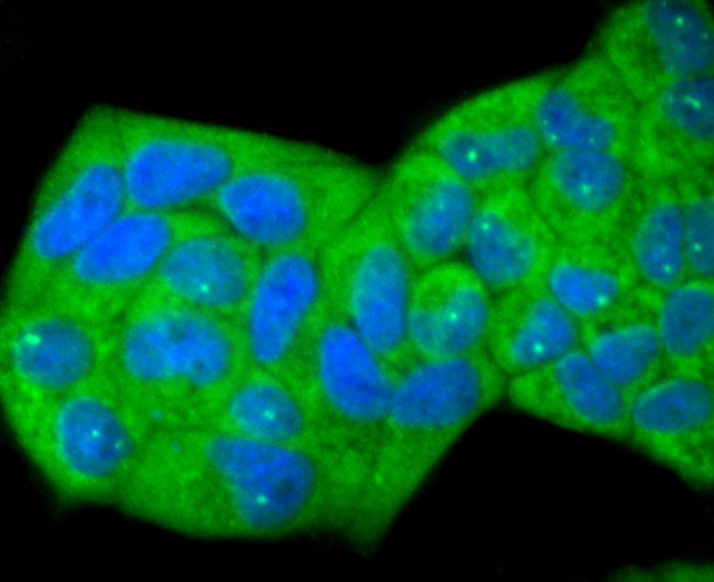
Fig3: ICC staining Id1 in Hela cells (green). The nuclear counter stain is DAPI (blue). Cells were fixed in paraformaldehyde, permeabilised with 0.25% Triton X100/PBS.Application
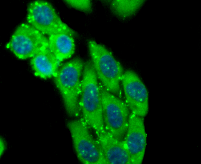
Fig4: ICC staining Id1 in HepG2 cells (green). The nuclear counter stain is DAPI (blue). Cells were fixed in paraformaldehyde, permeabilised with 0.25% Triton X100/PBS.Application
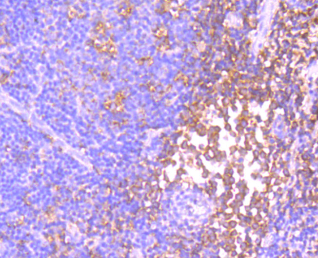
Fig5: Immunohistochemical analysis of paraffin-embedded human tonsil tissue using anti-Id1 antibody. Counter stained with hematoxylin.Application
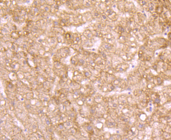
Fig6: Immunohistochemical analysis of paraffin-embedded human liver tissue using anti-Id1 antibody. Counter stained with hematoxylin.Application
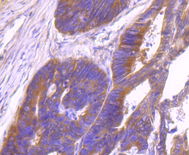
Fig7: Immunohistochemical analysis of paraffin-embedded human colon tissue using anti-Id1 antibody. Counter stained with hematoxylin.Application
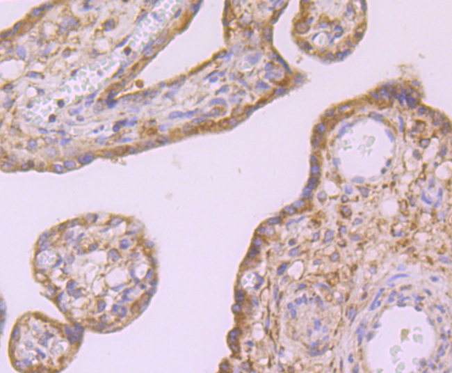
Fig8: Immunohistochemical analysis of paraffin-embedded human placenta tissue using anti-Id1 antibody. Counter stained with hematoxylin.Application
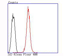
Fig9: Flow cytometric analysis of HepG2 cells with Id1 antibody at 1/100 dilution (red) compared with an unlabelled control (cells without incubation with primary antibody; black).| Product Name | Id1 Recombinant Rabbit Monoclonal Antibody |
|---|---|
| Antibody Type | Primary Antibodies |
| Product description | Members of the Id family of basic helix-loop-helix (bHLH) proteins include Id1, Id2, Id3 and Id4. They are ubiquitously expressed and dimerize with members of the class A and B HLH proteins. Due to the absence of the basic region, the resulting heterodimers cannot bind DNA. The Id-type proteins thus appear to negatively regulate DNA binding of bHLH proteins. Since Id1 inhibits DNA binding of E12 and Myo D, it apparently functions to inhibit muscle-specific gene expression. Under conditions that facilitate muscle cell differentiation, the Id protein levels fall, allowing E12 and/or E47 to form heterodimers with Myo D and myogenin, which in turn activate myogenic differentiation. It has been shown that expression of each of the Id proteins is strongly dependent on growth factor activation and that reduction of Id mRNA levels by antisense oligonucleotides leads to a delayed reentry of arrested cells into the cell cycle following growth factor stimulation. |
| Immunogen | Synthetic peptide within Human Id1 aa 11-60 / 155. |
| Clonality | Monoclonal |
|---|---|
| Isotype | IgG |
| Host Species | Recombinant rabbit |
| Tested Applications | FCICC/IFIHCWB |
| WB:1:500-1:2,000 ICC/IF:1:50-1:200 IHC:1:50-1:200 FC:1:50-1:100 |
|
| Species Reactivity | Human |
| Concentration | 1mg/ml |
| Alternative Names | bHLHb24 antibody
Class B basic helix-loop-helix protein 24 antibody
dJ857M17.1.2 (inhibitor of DNA binding 1 dominant negative helix-loop-helix protein) antibody DNA binding protein inhibitor ID 1 antibody DNA binding protein inhibitor ID1 antibody DNA-binding protein inhibitor ID-1 antibody Dominant negative helix loop helix protein antibody ID 1 antibody ID antibody ID1 antibody ID1_HUMAN antibody Inhibitor of Differentiation 1 antibody Inhibitor of DNA binding 1 antibody inhibitor of DNA binding 1 dominant negative helix-loop-helix protein antibody |
|---|---|
| Molecular Weight(MW) | 22 kDa |
| Cellular Localization | Cytoplasm. Nucleus. |
| SwissProt ID | P41134 |
|---|
Application
Fig1: Western blot analysis of Id1 on 293T cell using anti-Id1 antibody at 1/1,000 dilution.
Application
Fig2: Western blot analysis of Id1 on different lysates with Rabbit anti-Id1 antibody at 1/500 dilution. Lane 1: Hela cell lysate, Lane 2: HepG2 cell lysate, Lysates/proteins at 10 µg/Lane. Exposure time: 1 minute; 15% SDS-PAGE gel. Proteins were transferred to a PVDF membrane and blocked with 5% NFDM/TBST for 1 hour at room temperature. The primary antibody at 1/500 dilution was used in 5% NFDM/TBST at room temperature for 2 hours. Goat Anti-Rabbit IgG - HRP Secondary Antibody at 1:300,000 dilution was used for 1 hour at room temperature.
Application
Fig3: ICC staining Id1 in Hela cells (green). The nuclear counter stain is DAPI (blue). Cells were fixed in paraformaldehyde, permeabilised with 0.25% Triton X100/PBS.
Application
Fig4: ICC staining Id1 in HepG2 cells (green). The nuclear counter stain is DAPI (blue). Cells were fixed in paraformaldehyde, permeabilised with 0.25% Triton X100/PBS.
Application
Fig5: Immunohistochemical analysis of paraffin-embedded human tonsil tissue using anti-Id1 antibody. Counter stained with hematoxylin.
Application
Fig6: Immunohistochemical analysis of paraffin-embedded human liver tissue using anti-Id1 antibody. Counter stained with hematoxylin.
Application
Fig7: Immunohistochemical analysis of paraffin-embedded human colon tissue using anti-Id1 antibody. Counter stained with hematoxylin.
Application
Fig8: Immunohistochemical analysis of paraffin-embedded human placenta tissue using anti-Id1 antibody. Counter stained with hematoxylin.
Application
Fig9: Flow cytometric analysis of HepG2 cells with Id1 antibody at 1/100 dilution (red) compared with an unlabelled control (cells without incubation with primary antibody; black).| Application Notes | WB:1:500-1:2,000 ICC/IF:1:50-1:200 IHC:1:50-1:200 FC:1:50-1:100 |
|---|
| Form | Liquid |
|---|---|
| Storage Instructions | Store at +4℃ after thawing. Aliquot store at -20℃ or -80℃. Avoid repeated freeze / thaw cycles. |
| Storage Buffer | 1*TBS (pH7.4), 1%BSA, 40%Glycerol. Preservative: 0.05% Sodium Azide. |


