Application
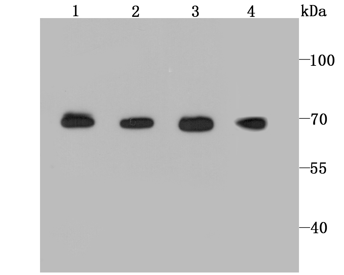
Fig1: Western blot analysis of BMAL1 on different lysates using anti-BMAL1 antibody at 1/1,000 dilution. Positive control: Lane 1: NIH/3T3 Lane 2: Rat brain Lane 3: Mouse spleen Lane 4: HelaApplication
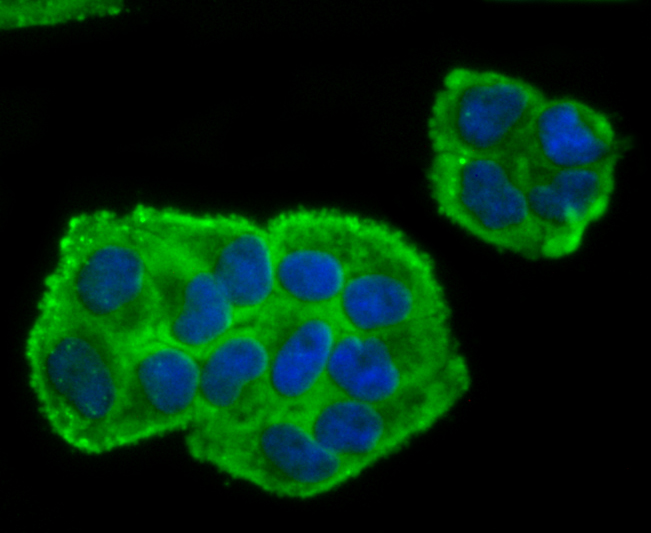
Fig2: ICC staining BMAL1 in Hela cells (green). The nuclear counter stain is DAPI (blue). Cells were fixed in paraformaldehyde, permeabilised with 0.25% Triton X100/PBS.Application
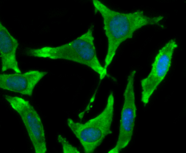
Fig3: ICC staining BMAL1 in SH-SY5Y cells (green). The nuclear counter stain is DAPI (blue). Cells were fixed in paraformaldehyde, permeabilised with 0.25% Triton X100/PBS.| Product Name | BMAL1 Recombinant Rabbit Monoclonal Antibody |
|---|---|
| Antibody Type | Primary Antibodies |
| Product description | Transcriptional activator which forms a core component of the circadian clock. Transcription factors, CLOCK or NPAS2 and ARNTL/BMAL1 or ARNTL2/BMAL2, form the positive limb of the feedback loop, act in the form of a heterodimer and activate the transcription of core clock genes and clock-controlled genes (involved in key metabolic processes), harboring E-box elements (5'-CACGTG-3') within their promoters. The core clock genes: PER1/2/3 and CRY1/2 which are transcriptional repressors form the negative limb of the feedback loop and interact with the CLOCK|NPAS2-ARNTL/BMAL1|ARNTL2/BMAL2 heterodimer inhibiting its activity and thereby negatively regulating their own expression. This heterodimer also activates nuclear receptors NR1D1/2 and RORA/B/G, which form a second feedback loop and which activate and repress ARNTL/BMAL1 transcription, respectively. ARNTL/BMAL1 positively regulates myogenesis and negatively regulates adipogenesis via the transcriptional control of the genes of the canonical Wnt signaling pathway. |
| Immunogen | recombinant protein |
| Clonality | Monoclonal |
|---|---|
| Isotype | IgG |
| Host Species | Recombinant rabbit |
| Tested Applications | ICC/IFWB |
| WB:1:1,000-1:2,000 ICC:1:50-1:200 |
|
| Species Reactivity | HumanMouseRat |
| Concentration | 1mg/ml |
| Alternative Names | ARNT like protein 1 brain and muscle antibody Arntl antibody Aryl hydrocarbon receptor nuclear translocator like antibody Aryl hydrocarbon receptor nuclear translocator like protein 1 antibody Aryl hydrocarbon receptor nuclear translocator-like protein 1 antibody Basic helix loop helix PAS orphan MOP3 antibody Basic helix loop helix PAS protein MOP3 antibody Basic-helix-loop-helix-PAS protein MOP3 antibody bHLH PAS protein JAP3 antibody bHLH-PAS protein JAP3 antibody bHLHe5 antibody BMAL 1 antibody BMAL1_HUMAN antibody BMAL1c antibody Brain and muscle ARNT like 1 antibody Brain and muscle ARNT-like 1 antibody CG8727 PA antibody Class E basic helix-loop-helix protein 5 antibody cycle antibody JAP 3 antibody JAP3 antibody Member of PAS protein 3 antibody Member of PAS superfamily 3 antibody MGC47515 antibody MOP 3 antibody MOP3 antibody PAS domain-containing protein 3 antibody PASD 3 antibody PASD3 antibody TIC antibody |
|---|---|
| Molecular Weight(MW) | 68 kDa |
| Cellular Localization | Nucleus. Cytoplasm. |
Application
Fig1: Western blot analysis of BMAL1 on different lysates using anti-BMAL1 antibody at 1/1,000 dilution. Positive control: Lane 1: NIH/3T3 Lane 2: Rat brain Lane 3: Mouse spleen Lane 4: Hela
Application
Fig2: ICC staining BMAL1 in Hela cells (green). The nuclear counter stain is DAPI (blue). Cells were fixed in paraformaldehyde, permeabilised with 0.25% Triton X100/PBS.
Application
Fig3: ICC staining BMAL1 in SH-SY5Y cells (green). The nuclear counter stain is DAPI (blue). Cells were fixed in paraformaldehyde, permeabilised with 0.25% Triton X100/PBS.| Positive Control | NIH/3T3, Hela, rat brain lysates, mouse spleen lysates, SH-SY5Y. |
|---|---|
| Application Notes | WB:1:1,000-1:2,000 ICC:1:50-1:200 |
| Form | Liquid |
|---|---|
| Storage Instructions | Store at +4℃ after thawing. Aliquot store at -20℃ or -80℃. Avoid repeated freeze / thaw cycles. |
| Storage Buffer | 1*TBS (pH7.4), 1%BSA, 40%Glycerol. Preservative: 0.05% Sodium Azide. |


