Application
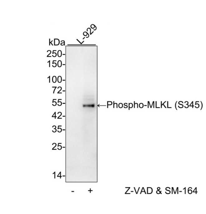
Fig1: Western blot analysis of Phospho-MLKL (S345) on different lysates with Rabbit anti-Phospho-MLKL (S345) antibody at 1/1,000 dilution. Lane 1: L-929 cell lysate, Lane 2: L929 treated with 20μM Z-VAD for 3.5 hours, then added 100nM SM-164 and 20ng/ml TNF-α for 3 hours cell lysate. Lysates/proteins at 20 µg/Lane. Exposure time: 1 minute 10 seconds; 4-20% SDS-PAGE gel. Proteins were transferred to a PVDF membrane and blocked with 5% NFDM/TBST for 1 hour at room temperature. The primary antibody at 1/1,000 dilution was used in 5% NFDM/TBST at 4℃ overnight. Goat Anti-Rabbit IgG - HRP Secondary Antibody at 1/50,000 dilution was used for 1 hour at room temperature.Application
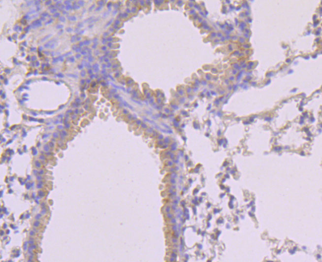
Fig2: Immunohistochemical analysis of paraffin-embedded mouse lung tissue using anti-MLKL (phospho S345) antibody. Counter stained with hematoxylin.Application
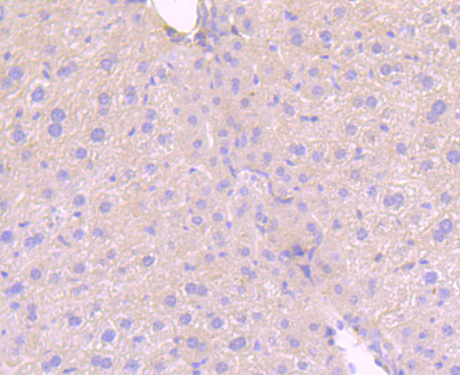
Fig3: Immunohistochemical analysis of paraffin-embedded mouse liver tissue using anti-MLKL (phospho S345) antibody. Counter stained with hematoxylin.Application
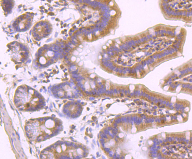
Fig4: Immunohistochemical analysis of paraffin-embedded mouse colon tissue using anti-MLKL (phospho S345) antibody. Counter stained with hematoxylin.Application
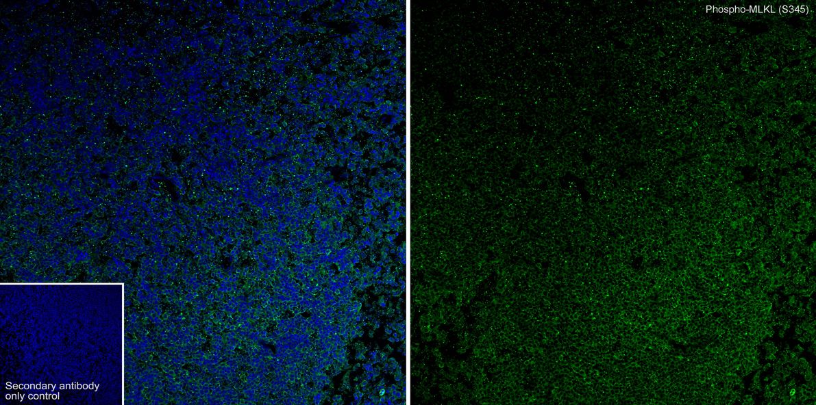
Fig5: Immunofluorescence analysis of frozen mouse spleen tissue with Rabbit anti-Phospho-MLKL (S345) antibody at 1/100 dilution. The section was pre-treated using heat mediated antigen retrieval with sodium citrate buffer (pH 6.0) for about 2 minutes in microwave oven. The tissues were blocked in 10% negative goat serum for 1 hour at room temperature, washed with PBS, and then probed with the primary antibody (green) at 1/100 dilution overnight at 4 ℃, washed with PBS. Goat Anti-Rabbit IgG H&L (488) was used as the secondary antibody at 1/1,000 dilution. Nuclei were counterstained with DAPI (blue).| Product Name | Phospho-MLKL (S345) Recombinant Rabbit Monoclonal Antibody |
|---|---|
| Antibody Type | Primary Antibodies |
| Product description | Pseudokinase that plays a key role in TNF-induced necroptosis, a programmed cell death process. Activated following phosphorylation by RIPK3, leading to homotrimerization, localization to the plasma membrane and execution of programmed necrosis characterized by calcium influx and plasma membrane damage. Does not have protein kinase activity. |
| Immunogen | Synthetic phospho-peptide corresponding to residues surrounding Ser345 of mouse MLKL aa 314-363 / 472. |
| Modification | p-S345 |
| Clonality | Monoclonal |
|---|---|
| Isotype | IgG |
| Host Species | Recombinant rabbit |
| Tested Applications | IF-FIHCWB |
| WB:1:1000 IHC:1:50-1:100 IF-F:1:100 |
|
| Species Reactivity | Mouse |
| Concentration | 1mg/ml |
| Purification | Protein A |
| Alternative Names | 9130019I15Rik antibody FLJ34389 antibody hMLKL antibody Mixed lineage kinase domain like antibody Mixed lineage kinase domain like protein antibody Mixed lineage kinase domain like pseudokinase antibody Mixed lineage kinase domain-like protein antibody Mlkl antibody MLKL_HUMAN antibody |
|---|---|
| Molecular Weight(MW) | 54kDa |
| Cellular Localization | Cell membrane, Cytoplasm, Nucleus. |
| SwissProt ID | Q9D2Y4 |
|---|
Application
Fig1: Western blot analysis of Phospho-MLKL (S345) on different lysates with Rabbit anti-Phospho-MLKL (S345) antibody at 1/1,000 dilution. Lane 1: L-929 cell lysate, Lane 2: L929 treated with 20μM Z-VAD for 3.5 hours, then added 100nM SM-164 and 20ng/ml TNF-α for 3 hours cell lysate. Lysates/proteins at 20 µg/Lane. Exposure time: 1 minute 10 seconds; 4-20% SDS-PAGE gel. Proteins were transferred to a PVDF membrane and blocked with 5% NFDM/TBST for 1 hour at room temperature. The primary antibody at 1/1,000 dilution was used in 5% NFDM/TBST at 4℃ overnight. Goat Anti-Rabbit IgG - HRP Secondary Antibody at 1/50,000 dilution was used for 1 hour at room temperature.
Application
Fig2: Immunohistochemical analysis of paraffin-embedded mouse lung tissue using anti-MLKL (phospho S345) antibody. Counter stained with hematoxylin.
Application
Fig3: Immunohistochemical analysis of paraffin-embedded mouse liver tissue using anti-MLKL (phospho S345) antibody. Counter stained with hematoxylin.
Application
Fig4: Immunohistochemical analysis of paraffin-embedded mouse colon tissue using anti-MLKL (phospho S345) antibody. Counter stained with hematoxylin.
Application
Fig5: Immunofluorescence analysis of frozen mouse spleen tissue with Rabbit anti-Phospho-MLKL (S345) antibody at 1/100 dilution. The section was pre-treated using heat mediated antigen retrieval with sodium citrate buffer (pH 6.0) for about 2 minutes in microwave oven. The tissues were blocked in 10% negative goat serum for 1 hour at room temperature, washed with PBS, and then probed with the primary antibody (green) at 1/100 dilution overnight at 4 ℃, washed with PBS. Goat Anti-Rabbit IgG H&L (488) was used as the secondary antibody at 1/1,000 dilution. Nuclei were counterstained with DAPI (blue).| Application Notes | WB:1:1000 IHC:1:50-1:100 IF-F:1:100 |
|---|
| Form | Liquid |
|---|---|
| Storage Instructions | Store at +4℃ after thawing. Aliquot store at -20℃ or -80℃. Avoid repeated freeze / thaw cycles. |
| Storage Buffer | 1*TBS (pH7.4), 1%BSA, 40%Glycerol. Preservative: 0.05% Sodium Azide. |


