Application
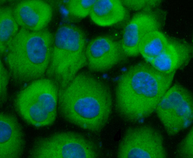
Fig1: ICC staining Carcino Embryonic Antigen CEA(CD66e) in HUVEC cells (green). The nuclear counter stain is DAPI (blue). Cells were fixed in paraformaldehyde, permeabilised with 0.25% Triton X100/PBS.Application
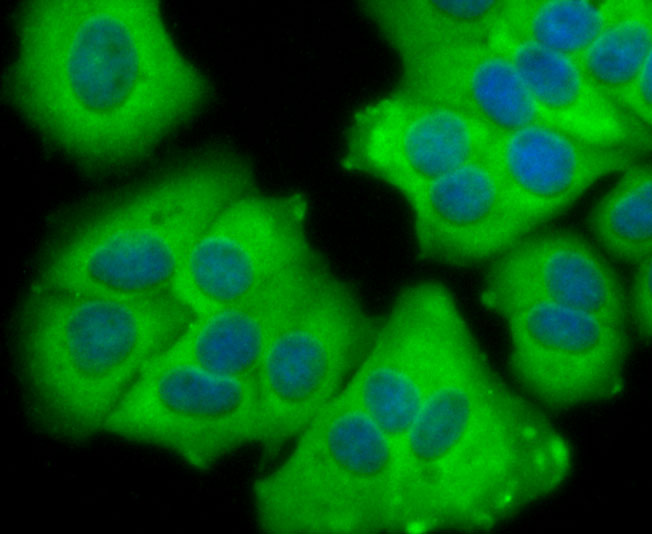
Fig2: ICC staining Carcino Embryonic Antigen CEA(CD66e) in MCF-7 cells (green). The nuclear counter stain is DAPI (blue). Cells were fixed in paraformaldehyde, permeabilised with 0.25% Triton X100/PBS.Application
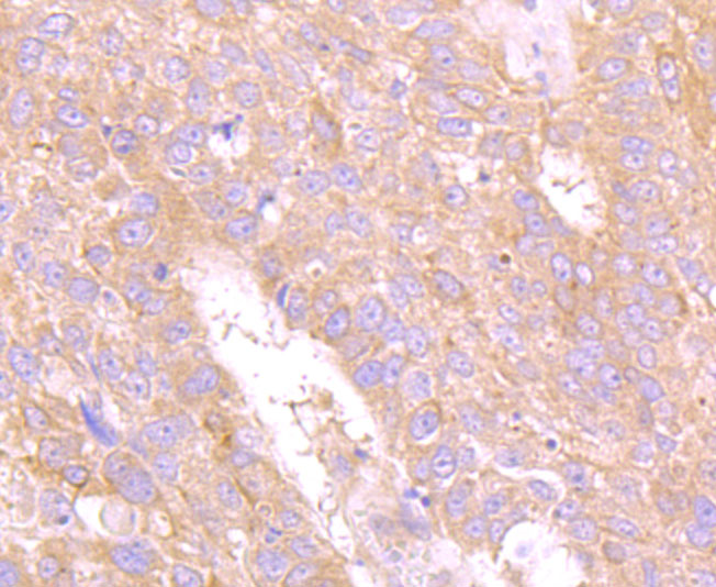
Fig3: Immunohistochemical analysis of paraffin-embedded human lung cancer tissue using anti-Carcino Embryonic Antigen CEA(CD66e) antibody. Counter stained with hematoxylin.Application
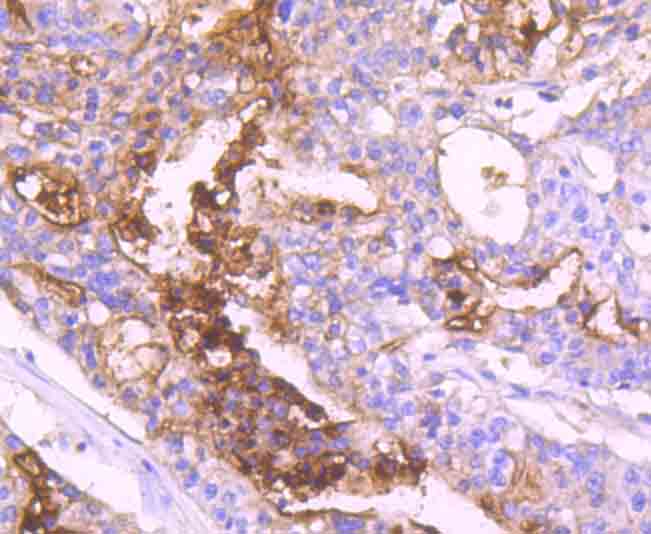
Fig4: Immunohistochemical analysis of paraffin-embedded human stomach cancer tissue using anti-Carcino Embryonic Antigen CEA(CD66e) antibody. Counter stained with hematoxylin.Application
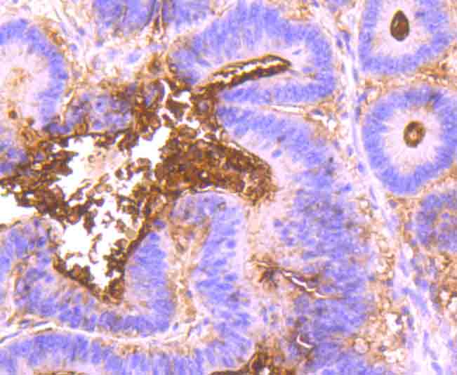
Fig5: Immunohistochemical analysis of paraffin-embedded human colon cancer tissue using anti-Carcino Embryonic Antigen CEA(CD66e) antibody. Counter stained with hematoxylin.| Product Name | Carcino Embryonic Antigen CEA [JM93-28] |
|---|---|
| Antibody Type | Primary Antibodies |
| Antigen Alias | Carcinoembryonic antigen antibody Carcinoembryonic antigen-related cell adhesion molecule 5 antibody CD66e antibody CEA antibody Ceacam5 antibody CEAM5_HUMAN antibody DKFZp781M2392 antibody Meconium antigen 100 antibody OTTHUMP00000199032 antibody OTTHUMP00000199033 antibody OTTHUMP00000199034 antibody |
| Product description | The CD66 (carcinoembryonic antigen, CEA, biliary glycoprotein I, BGP-1, CEACAM) immunoglobulin superfamily of genes encode cell adhesion proteins, which are expressed at higher levels in tumorous tissues than in normal tissues. The human CD66 gene family is a diverse set of glycoproteins of epithelial and hematopoietic lineage that comprises 29 genes, which map to chromosome position 19q13.1-q13.2. CD66A, CD66B, CD66C, CD66D, CD66E and CD66F are the best characterized CD66 antigens, and CD66A-D expression upregulates on the surface of granulocytes upon stimulation. Certain CD66 family members mediate homotypic and heterotypic intercellular adhesion events. CD66E, also known as CEA, is a well known tumor marker and a heavily glycosylated GPI-linked cell surface molecule. |
| Immunogen | Recombinant protein |
| Clonality | Monoclonal |
|---|---|
| Isotype | IgG |
| Host Species | Recombinant rabbit |
| Tested Applications | WBICC/IFIHC |
| WB:1:500 ICC:1:50-1:200 IHC:1:50-1:200 |
|
| Species Reactivity | Human |
| Concentration | 1mg/ml |
| Alternative Names | Carcinoembryonic antigen antibody Carcinoembryonic antigen-related cell adhesion molecule 5 antibody CD66e antibody CEA antibody Ceacam5 antibody CEAM5_HUMAN antibody DKFZp781M2392 antibody Meconium antigen 100 antibody OTTHUMP00000199032 antibody OTTHUMP00000199033 antibody OTTHUMP00000199034 antibody |
|---|---|
| Molecular Weight(MW) | 76 kDa |
| Cellular Localization | Membrane. |
| SwissProt ID | P06731 |
|---|
Application
Fig1: ICC staining Carcino Embryonic Antigen CEA(CD66e) in HUVEC cells (green). The nuclear counter stain is DAPI (blue). Cells were fixed in paraformaldehyde, permeabilised with 0.25% Triton X100/PBS.
Application
Fig2: ICC staining Carcino Embryonic Antigen CEA(CD66e) in MCF-7 cells (green). The nuclear counter stain is DAPI (blue). Cells were fixed in paraformaldehyde, permeabilised with 0.25% Triton X100/PBS.
Application
Fig3: Immunohistochemical analysis of paraffin-embedded human lung cancer tissue using anti-Carcino Embryonic Antigen CEA(CD66e) antibody. Counter stained with hematoxylin.
Application
Fig4: Immunohistochemical analysis of paraffin-embedded human stomach cancer tissue using anti-Carcino Embryonic Antigen CEA(CD66e) antibody. Counter stained with hematoxylin.
Application
Fig5: Immunohistochemical analysis of paraffin-embedded human colon cancer tissue using anti-Carcino Embryonic Antigen CEA(CD66e) antibody. Counter stained with hematoxylin.| Positive Control | HUVEC, MCF-7, human lung cancer tissue, human stomach cancer tissue, human colon cancer tissue. |
|---|---|
| Application Notes | WB:1:500 ICC:1:50-1:200 IHC:1:50-1:200 |
| Form | Liquid |
|---|---|
| Storage Instructions | Store at +4℃ after thawing. Aliquot store at -20℃ or -80℃. Avoid repeated freeze / thaw cycles. |
| Storage Buffer | 1*TBS (pH7.4), 1%BSA, 40%Glycerol. Preservative: 0.05% Sodium Azide. |


