WB
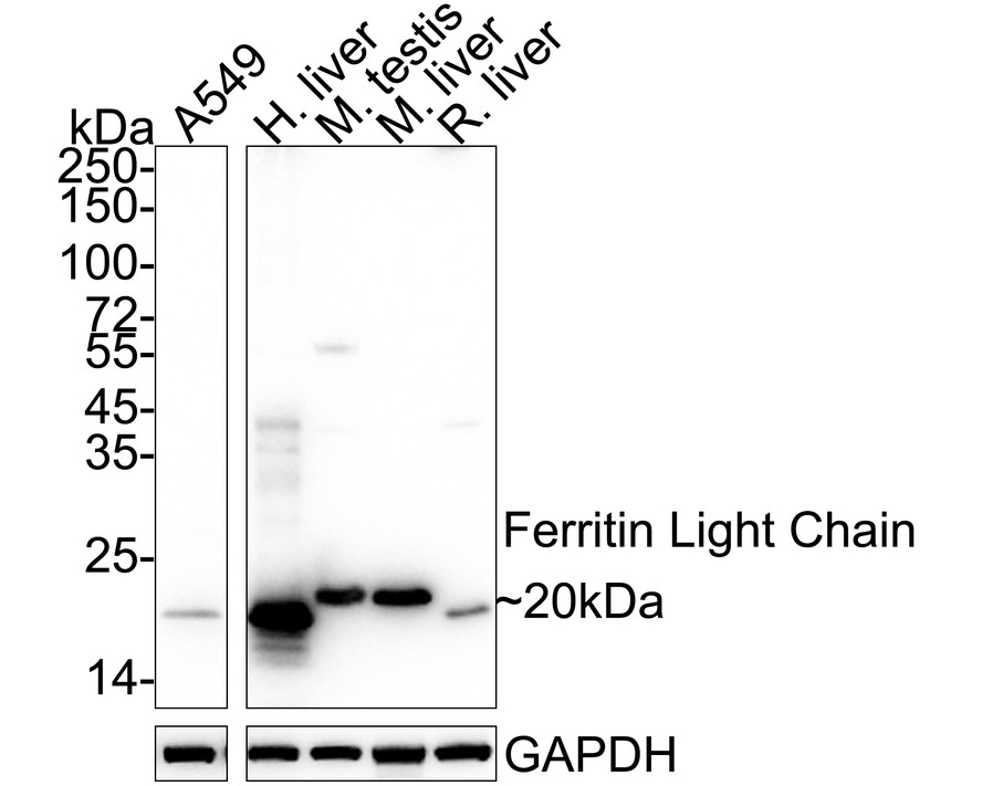
Western blot analysis of Ferritin Light Chain on different lysates with Rabbit anti-Ferritin Light Chain antibody at 1/1,000 dilution. Lane 1: A549 cell lysate (20 µg/Lane), Lane 2: Human liver tissue lysate (40 µg/Lane), Lane 3: Mouse testis tissue lysate (40 µg/Lane), Lane 4: Mouse liver tissue lysate (40 µg/Lane), Lane 5: Rat liver tissue lysate (40 µg/Lane), Exposure time: 2 seconds; 4-20% SDS-PAGE gel. Proteins were transferred to a PVDF membrane and blocked with 5% NFDM/TBST for 1 hour at room temperature. The primary antibody at 1/1,000 dilution was used in 5% NFDM/TBST at 4℃ overnight. Goat Anti-Rabbit IgG - HRP Secondary Antibody at 1/50,000 dilution was used for 1 hour at room temperature.IHC
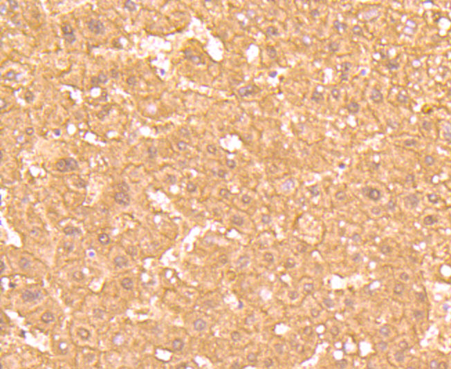
Immunohistochemical analysis of paraffin-embedded human liver tissue using anti-Ferritin Light Chain antibody. Counter stained with hematoxylin.IHC
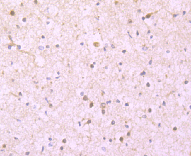
Immunohistochemical analysis of paraffin-embedded human brain tissue using anti-Ferritin Light Chain antibody. Counter stained with hematoxylin.IHC
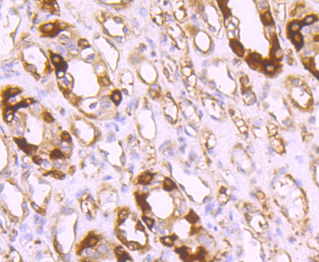
Immunohistochemical analysis of paraffin-embedded human kidney tissue using anti-Ferritin Light Chain antibody. Counter stained with hematoxylin.IHC
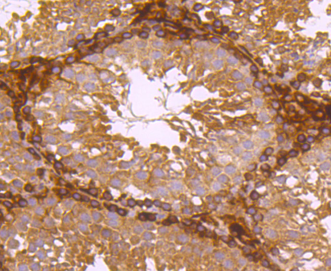
Immunohistochemical analysis of paraffin-embedded mouse testes tissue using anti-Ferritin Light Chain antibody. Counter stained with hematoxylin.IHC
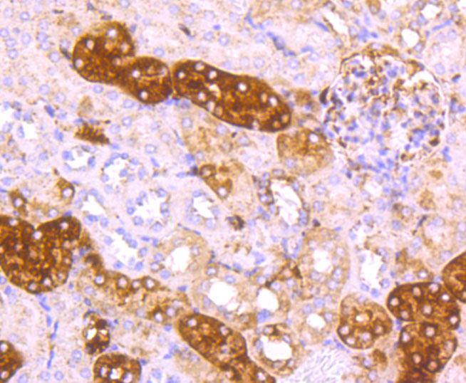
Immunohistochemical analysis of paraffin-embedded mouse kidney tissue using anti-Ferritin Light Chain antibody. Counter stained with hematoxylin.| Product Name | Ferritin Light Chain Recombinant Rabbit Monoclonal Antibody |
|---|---|
| Antibody Type | Primary Antibodies |
| Product description | Mammalian ferritins consist of 24 subunits made up of two types of polypeptide chains, ferritin heavy chain and ferritin light chain, which each have unique functions. Ferritin heavy chains catalyze the first step in iron storage, the oxidation of Fe (II), whereas ferritin light chains promote the nucleation of ferrihydrite, enabling storage of Fe (III). The most prominent role of mammalian ferritins is to provide iron-buffering capacity to cells. In addition to iron buffering, heavy chain ferritin is also involved in the regulation of thymidine biosynthesis via increased expression of cytoplasmic serine hydroxymethyltransferase, which is a limiting factor in thymidylate synthesis in MCF-7 cells. Light chain ferritin is involved in cataracts by at least two mechanisms, hereditary hyperferritinemia cataract syndrome, in which light chain ferritin is overexpressed, and oxidative stress, an important factor in the development of ageing-related cataracts. The gene encoding human ferritin heavy chain maps to chromosome 11q13 and the human ferritin light chain gene maps to chromosome 19q13.3-q13.4. |
| Immunogen | Synthetic peptide within Human Ferritin Light Chain aa 126-175 / 175. |
| Clonality | Monoclonal |
|---|---|
| Isotype | IgG |
| Host Species | Recombinant rabbit |
| Tested Applications | IHCWB |
| WB:1:1000-1:5000 IHC:1:100-1:500 |
|
| Species Reactivity | HumanMouseRat |
| Concentration | 1mg/ml |
| Purification | Protein A |
| Alternative Names | Ferritin L chain antibody Ferritin L subunit antibody Ferritin light chain antibody Ferritin light polypeptide antibody ferritin light polypeptide like 3 antibody FRIL_HUMAN antibody FTL antibody LFTD antibody NBIA 3 antibody NBIA3 antibody |
|---|---|
| Molecular Weight(MW) | 20 kDa |
| Cellular Localization | Cytoplasm. Membrane. Nucleus. |
WB
Western blot analysis of Ferritin Light Chain on different lysates with Rabbit anti-Ferritin Light Chain antibody at 1/1,000 dilution. Lane 1: A549 cell lysate (20 µg/Lane), Lane 2: Human liver tissue lysate (40 µg/Lane), Lane 3: Mouse testis tissue lysate (40 µg/Lane), Lane 4: Mouse liver tissue lysate (40 µg/Lane), Lane 5: Rat liver tissue lysate (40 µg/Lane), Exposure time: 2 seconds; 4-20% SDS-PAGE gel. Proteins were transferred to a PVDF membrane and blocked with 5% NFDM/TBST for 1 hour at room temperature. The primary antibody at 1/1,000 dilution was used in 5% NFDM/TBST at 4℃ overnight. Goat Anti-Rabbit IgG - HRP Secondary Antibody at 1/50,000 dilution was used for 1 hour at room temperature.
IHC
Immunohistochemical analysis of paraffin-embedded human liver tissue using anti-Ferritin Light Chain antibody. Counter stained with hematoxylin.
IHC
Immunohistochemical analysis of paraffin-embedded human brain tissue using anti-Ferritin Light Chain antibody. Counter stained with hematoxylin.
IHC
Immunohistochemical analysis of paraffin-embedded human kidney tissue using anti-Ferritin Light Chain antibody. Counter stained with hematoxylin.
IHC
Immunohistochemical analysis of paraffin-embedded mouse testes tissue using anti-Ferritin Light Chain antibody. Counter stained with hematoxylin.
IHC
Immunohistochemical analysis of paraffin-embedded mouse kidney tissue using anti-Ferritin Light Chain antibody. Counter stained with hematoxylin.| Application Notes | WB:1:1000-1:5000 IHC:1:100-1:500 |
|---|
| Form | Liquid |
|---|---|
| Storage Instructions | Store at +4℃ after thawing. Aliquot store at -20℃ or -80℃. Avoid repeated freeze / thaw cycles. |
| Storage Buffer | 1*TBS (pH7.4), 1%BSA, 40%Glycerol. Preservative: 0.05% Sodium Azide. |


