WB
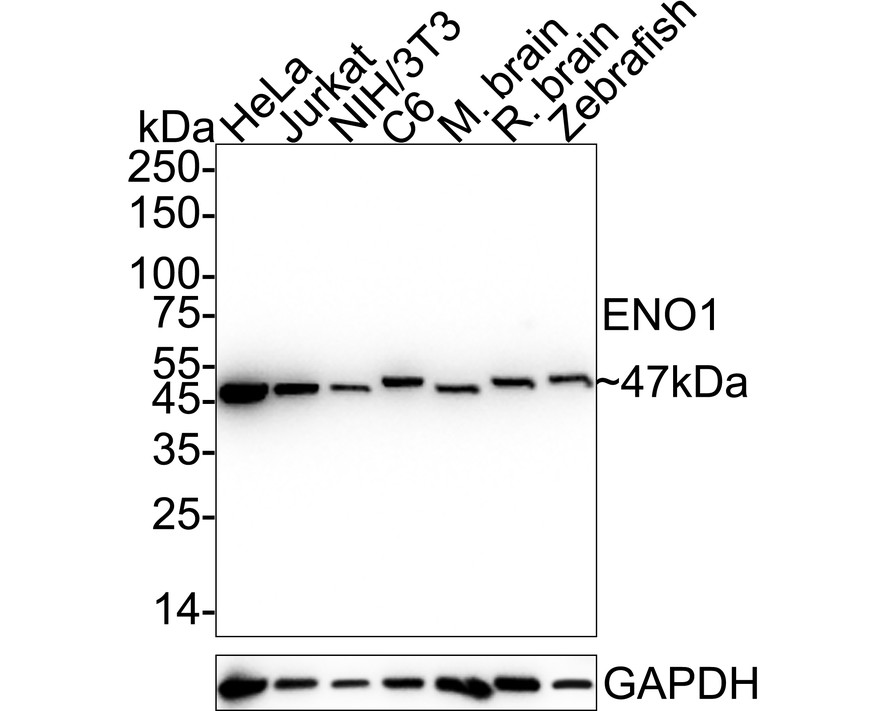
Western blot analysis of ENO1 on different lysates with Rabbit anti-ENO1 antibody at 1/2,000 dilution. Lane 1: HeLa cell lysate (20 µg/Lane), Lane 2: Jurkat cell lysate (20 µg/Lane), Lane 3: NIH/3T3 cell lysate (20 µg/Lane), Lane 4: C6 cell lysate (20 µg/Lane), Lane 5: Mouse brain tissue lysate (20 µg/Lane), Lane 6: Rat brain tissue lysate (20 µg/Lane), Lane 7: Zebrafish tissue lysate (20 µg/Lane), Exposure time: 6 seconds; 4-20% SDS-PAGE gel. Proteins were transferred to a PVDF membrane and blocked with 5% NFDM/TBST for 1 hour at room temperature. The primary antibody at 1/2,000 dilution was used in 5% NFDM/TBST at 4℃ overnight. Goat Anti-Rabbit IgG - HRP Secondary Antibody at 1/50,000 dilution was used for 1 hour at room temperature.IHC
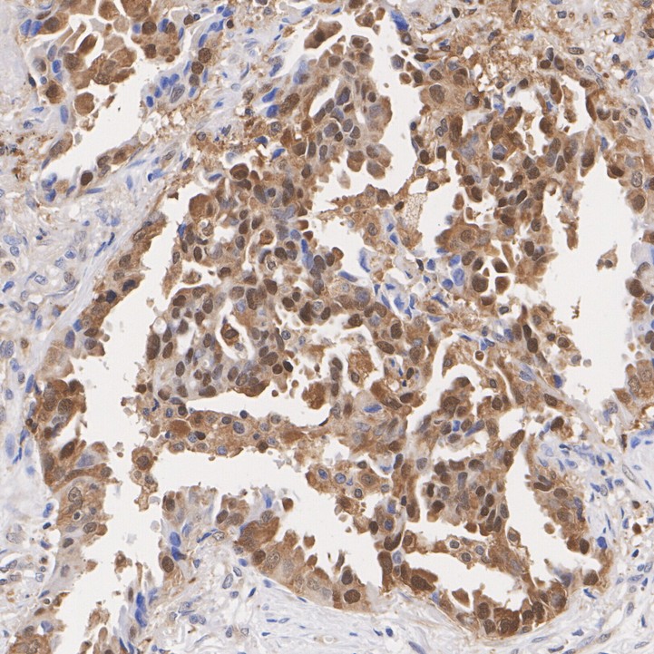
Immunohistochemical analysis of paraffin-embedded human lung cancer tissue with Rabbit anti-ENO1 antibody at 1/1,000 dilution. The section was pre-treated using heat mediated antigen retrieval with Tris-EDTA buffer (pH 9.0) for 20 minutes. The tissues were blocked in 1% BSA for 20 minutes at room temperature, washed with ddH2O and PBS, and then probed with the primary antibody at 1/1,000 dilution for 1 hour at room temperature. The detection was performed using an HRP conjugated compact polymer system. DAB was used as the chromogen. Tissues were counterstained with hematoxylin and mounted with DPX.ICC/IF
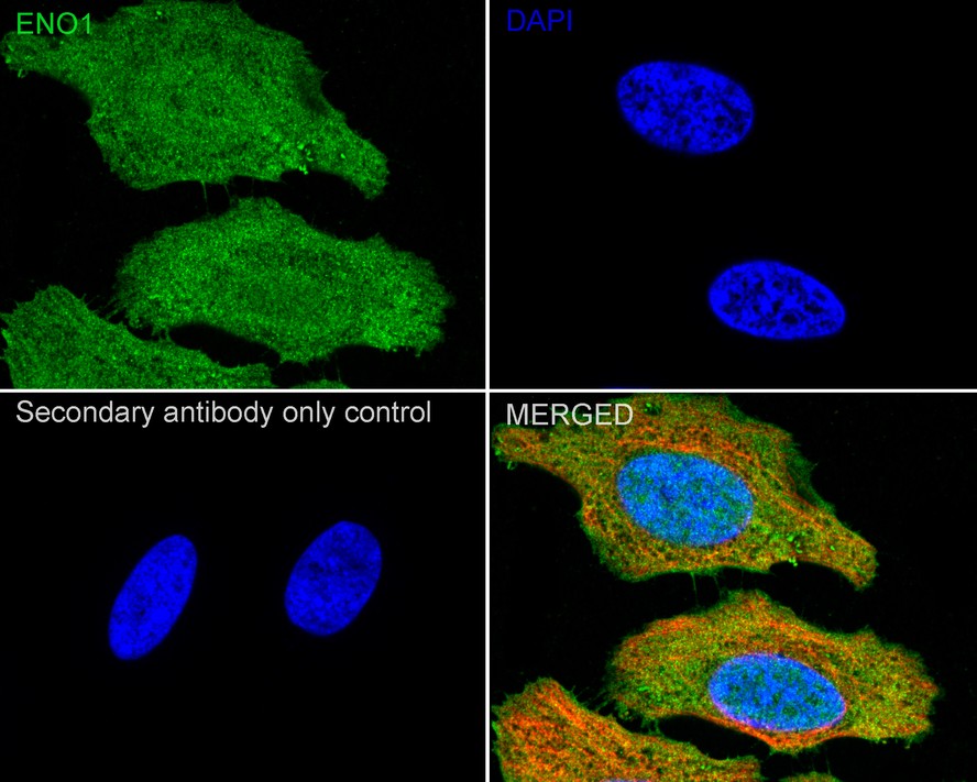
Immunocytochemistry analysis of HeLa cells labeling ENO1 with Rabbit anti-ENO1 antibody at 1/100 dilution. Cells were fixed in 4% paraformaldehyde for 15 minutes at room temperature, permeabilized with 0.1% Triton X-100 in PBS for 15 minutes at room temperature, then blocked with 1% BSA in 10% negative goat serum for 1 hour at room temperature. Cells were then incubated with Rabbit anti-ENO1 antibody at 1/100 dilution in 1% BSA in PBST overnight at 4 ℃. Goat Anti-Rabbit IgG H&L (488) was used as the secondary antibody at 1/1,000 dilution. PBS instead of the primary antibody was used as the secondary antibody only control. Nuclear DNA was labelled in blue with DAPI. Beta tubulin (red) was stained at 1/100 dilution overnight at +4℃. Goat Anti-Mouse IgG H&L (594) was used as the secondary antibody at 1/1,000 dilution.FC
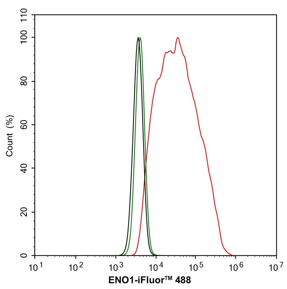
Flow cytometric analysis of HeLa cells labeling ENO1. Cells were fixed and permeabilized. Then stained with the primary antibody (1/1,000) (red) compared with Rabbit IgG Isotype Control (green). After incubation of the primary antibody at +4℃ for an hour, the cells were stained with a 488 conjugate-Goat anti-Rabbit IgG Secondary antibody at 1/1,000 dilution for 30 minutes at +4℃. Unlabelled sample was used as a control (cells without incubation with primary antibody; black).| Product Name | ENO1 Recombinant Rabbit Monoclonal Antibody |
|---|---|
| Antibody Type | Primary Antibodies |
| Product description | Multifunctional enzyme that, as well as its role in glycolysis, plays a part in various processes such as growth control, hypoxia tolerance and allergic responses. May also function in the intravascular and pericellular fibrinolytic system due to its ability to serve as a receptor and activator of plasminogen on the cell surface of several cell-types such as leukocytes and neurons. Stimulates immunoglobulin production. MBP1 binds to the myc promoter and acts as a transcriptional repressor. May be a tumor suppressor. |
| Immunogen | Synthetic peptide within Human ENO1 aa 1-50 / 434. |
| Clonality | Monoclonal |
|---|---|
| Isotype | IgG |
| Host Species | Recombinant rabbit |
| Tested Applications | FCICC/IFIHCWB |
| WB:1:2000 IHC:1:1000 ICC/IF:1:100 FC:1:1000 |
|
| Species Reactivity | HumanMouseRatZebrafish |
| Concentration | 1mg/ml |
| Purification | Protein A |
| Alternative Names | 2 phospho D glycerate hydro lyase antibody 2-phospho-D-glycerate hydro-lyase antibody Alpha enolase antibody Alpha enolase like 1 antibody Alpha-enolase antibody C myc promoter binding protein antibody C-myc promoter-binding protein antibody EC 4.2.1.11 antibody eno1 antibody ENO1L1 antibody ENOA_HUMAN antibody Enolase 1 (alpha) antibody Enolase 1 (alpha) like 1 antibody Enolase 1 antibody Enolase alpha antibody MBP 1 antibody MBP-1 antibody MBP1 antibody MBPB1 antibody MPB 1 antibody MPB-1 antibody MPB1 antibody MYC promoter binding protein 1 antibody NNE antibody Non neural enolase antibody Non-neural enolase antibody Phosphopyruvate hydratase antibody Plasminogen binding protein antibody Plasminogen-binding protein antibody PPH antibody Tau crystallin antibody |
|---|---|
| Molecular Weight(MW) | 47 kDa |
| Cellular Localization | Cell membrane, Cytoplasm, Membrane, Nucleus. |
WB
Western blot analysis of ENO1 on different lysates with Rabbit anti-ENO1 antibody at 1/2,000 dilution. Lane 1: HeLa cell lysate (20 µg/Lane), Lane 2: Jurkat cell lysate (20 µg/Lane), Lane 3: NIH/3T3 cell lysate (20 µg/Lane), Lane 4: C6 cell lysate (20 µg/Lane), Lane 5: Mouse brain tissue lysate (20 µg/Lane), Lane 6: Rat brain tissue lysate (20 µg/Lane), Lane 7: Zebrafish tissue lysate (20 µg/Lane), Exposure time: 6 seconds; 4-20% SDS-PAGE gel. Proteins were transferred to a PVDF membrane and blocked with 5% NFDM/TBST for 1 hour at room temperature. The primary antibody at 1/2,000 dilution was used in 5% NFDM/TBST at 4℃ overnight. Goat Anti-Rabbit IgG - HRP Secondary Antibody at 1/50,000 dilution was used for 1 hour at room temperature.
IHC
Immunohistochemical analysis of paraffin-embedded human lung cancer tissue with Rabbit anti-ENO1 antibody at 1/1,000 dilution. The section was pre-treated using heat mediated antigen retrieval with Tris-EDTA buffer (pH 9.0) for 20 minutes. The tissues were blocked in 1% BSA for 20 minutes at room temperature, washed with ddH2O and PBS, and then probed with the primary antibody at 1/1,000 dilution for 1 hour at room temperature. The detection was performed using an HRP conjugated compact polymer system. DAB was used as the chromogen. Tissues were counterstained with hematoxylin and mounted with DPX.
ICC/IF
Immunocytochemistry analysis of HeLa cells labeling ENO1 with Rabbit anti-ENO1 antibody at 1/100 dilution. Cells were fixed in 4% paraformaldehyde for 15 minutes at room temperature, permeabilized with 0.1% Triton X-100 in PBS for 15 minutes at room temperature, then blocked with 1% BSA in 10% negative goat serum for 1 hour at room temperature. Cells were then incubated with Rabbit anti-ENO1 antibody at 1/100 dilution in 1% BSA in PBST overnight at 4 ℃. Goat Anti-Rabbit IgG H&L (488) was used as the secondary antibody at 1/1,000 dilution. PBS instead of the primary antibody was used as the secondary antibody only control. Nuclear DNA was labelled in blue with DAPI. Beta tubulin (red) was stained at 1/100 dilution overnight at +4℃. Goat Anti-Mouse IgG H&L (594) was used as the secondary antibody at 1/1,000 dilution.
FC
Flow cytometric analysis of HeLa cells labeling ENO1. Cells were fixed and permeabilized. Then stained with the primary antibody (1/1,000) (red) compared with Rabbit IgG Isotype Control (green). After incubation of the primary antibody at +4℃ for an hour, the cells were stained with a 488 conjugate-Goat anti-Rabbit IgG Secondary antibody at 1/1,000 dilution for 30 minutes at +4℃. Unlabelled sample was used as a control (cells without incubation with primary antibody; black).| Positive Control | . |
|---|---|
| Application Notes | WB:1:2000 IHC:1:1000 ICC/IF:1:100 FC:1:1000 |
| Form | Liquid |
|---|---|
| Storage Instructions | Store at +4℃ after thawing. Aliquot store at -20℃ or -80℃. Avoid repeated freeze / thaw cycles. |
| Storage Buffer | 1*TBS (pH7.4), 1%BSA, 40%Glycerol. Preservative: 0.05% Sodium Azide. |


